


 Foundational characteristics of cancer include proliferation, angiogenesis, migration, evasion of apoptosis, and cellular immortality. Find key markers for these cellular processes and antibodies to detect them.
 The SUMOplot™ Analysis Program predicts and scores sumoylation sites in your protein. SUMOylation is a post-translational modification involved in various cellular processes, such as nuclear-cytosolic transport, transcriptional regulation, apoptosis, protein stability, response to stress, and progression through the cell cycle.
 The Autophagy Receptor Motif Plotter predicts and scores autophagy receptor binding sites in your protein. Identifying proteins connected to this pathway is critical to understanding the role of autophagy in physiological as well as pathological processes such as development, differentiation, neurodegenerative diseases, stress, infection, and cancer.
> home > Products > Primary Antibodies > Antibody Collections > KD-Validated Antibodies > KD-Validated Anti-JUP Rabbit Monoclonal Antibody
KD-Validated Anti-JUP Rabbit Monoclonal Antibody
Rabbit monoclonal antibody
- SPECIFICATION
- CITATIONS
- PROTOCOLS
- BACKGROUND

Application
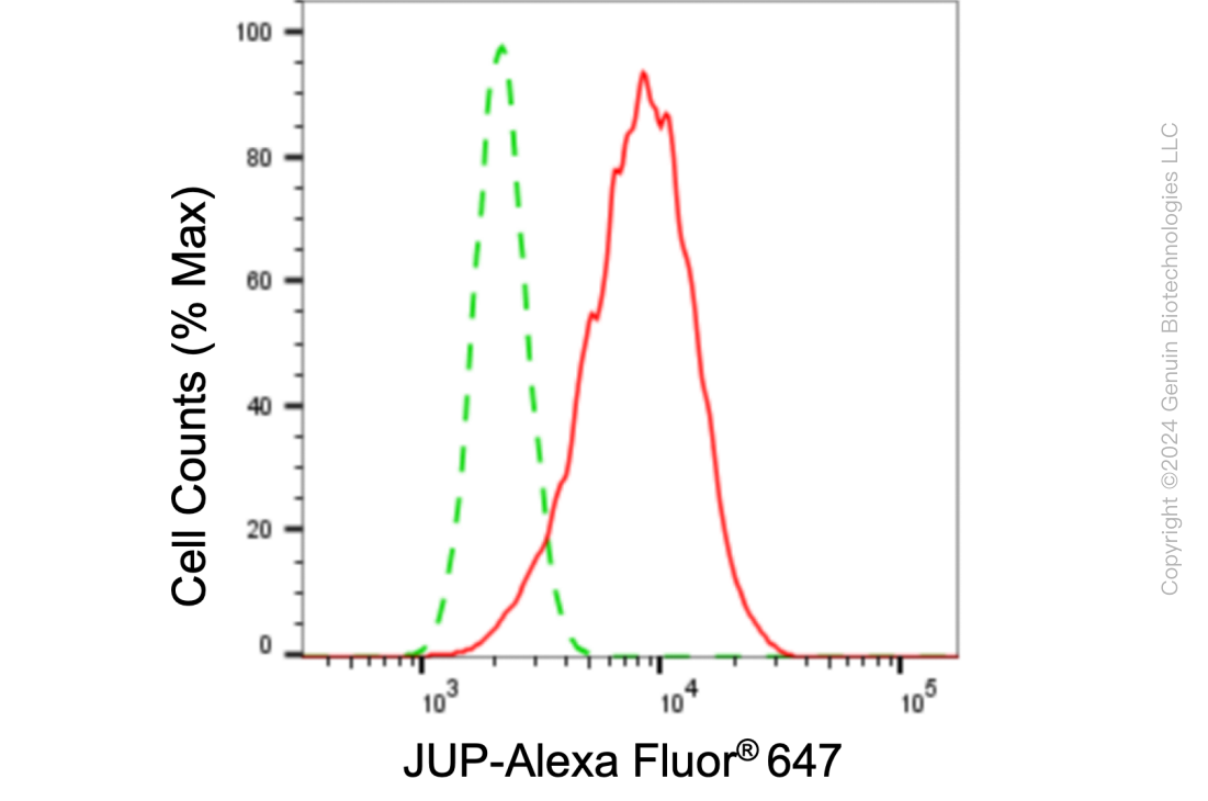
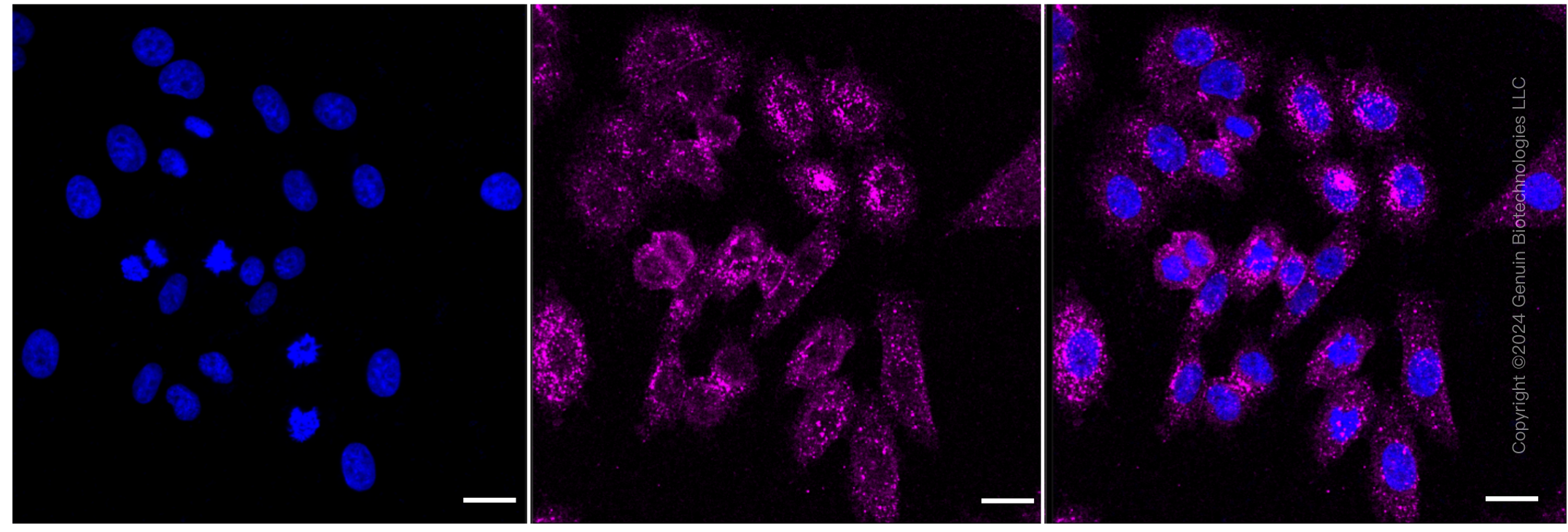
| WB, FC, ICC |
|---|---|
| Primary Accession | P14923 |
| Reactivity | Human, Mouse |
| Clonality | Monoclonal |
| Isotype | Rabbit IgG |
| Clone Names | 24GB6055 |
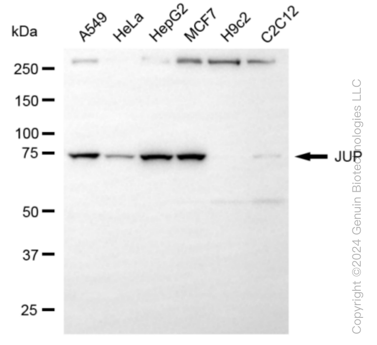
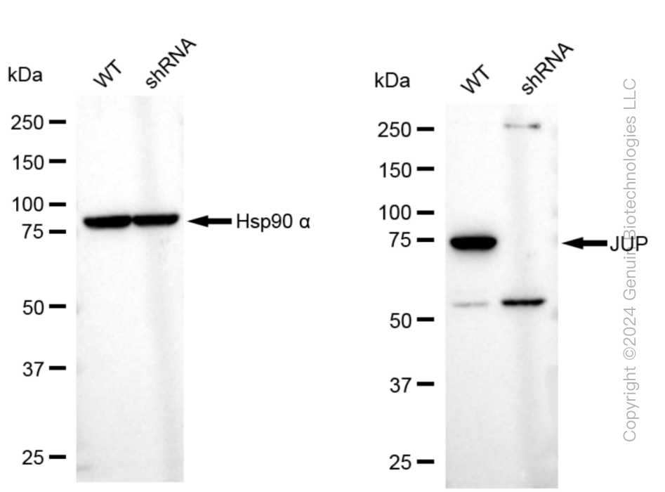
| Calculated MW | Predicted, 82 kDa, observed, 75 kDa |
| Gene Name | JUP |
| Aliases | JUP; Junction Plakoglobin; DP3; DPIII; CTNNG; PDGB; PKGB; PG; Catenin (Cadherin-Associated Protein), Gamma 80kDa; Desmosomal Protein 3; Desmoplakin III; Desmoplakin-3; Catenin Gamma |
| Immunogen | A synthesized peptide derived from human Catenin gamma |
| Gene ID | 3728 |
|---|---|
| Other Names | Junction plakoglobin, Catenin gamma, Desmoplakin III, Desmoplakin-3, JUP (HGNC:6207) |
| Name | JUP (HGNC:6207) |
|---|---|
| Function | Common junctional plaque protein. The membrane-associated plaques are architectural elements in an important strategic position to influence the arrangement and function of both the cytoskeleton and the cells within the tissue. The presence of plakoglobin in both the desmosomes and in the intermediate junctions suggests that it plays a central role in the structure and function of submembranous plaques. Acts as a substrate for VE-PTP and is required by it to stimulate VE- cadherin function in endothelial cells. Can replace beta-catenin in E- cadherin/catenin adhesion complexes which are proposed to couple cadherins to the actin cytoskeleton (By similarity). |
| Cellular Location | Cell junction, adherens junction. Cell junction, desmosome. Cytoplasm, cytoskeleton. Cell membrane; Peripheral membrane protein. Cytoplasm {ECO:0000250|UniProtKB:Q9PVF7}. Cell junction {ECO:0000250|UniProtKB:Q9PVF7}. Nucleus {ECO:0000250|UniProtKB:Q9PVF7} Note=Cytoplasmic in a soluble and membrane-associated form. Colocalizes with DSG4 at desmosomes (PubMed:21495994) |
| Tissue Location | Expressed in the heart (at protein level). |
Research Areas
Citations (0)

Thousands of laboratories across the world have published research that depended on the performance of antibodies from Abcepta to advance their research. Check out links to articles that cite our products in major peer-reviewed journals, organized by research category.
Submit your citation using an Abcepta antibody to
info@abcepta.com, and receive a free "I Love Antibodies" mug.
info@abcepta.com, and receive a free "I Love Antibodies" mug.

Application Protocols
Provided below are standard protocols that you may find useful for product applications.
Abcepta welcomes feedback from its customers.
If you have used an Abcepta product and would like to share how it has performed, please click on the "Submit Review" button and provide the requested information. Our staff will examine and post your review and contact you if needed.
If you have any additional inquiries please email technical services at tech@abcepta.com.
$ 399.20

$ 149.00
Cat# AGI1054
Ordering Information
United States
AlbaniaAustraliaAustriaBelgiumBosnia & HerzegovinaBrazilBulgariaCanadaCentral AmericaChinaCroatiaCyprusCzech RepublicDenmarkEstoniaFinlandFranceGermanyGreeceHong KongHungaryIcelandIndiaIndonesiaIrelandIsraelItalyJapanLatviaLithuaniaLuxembourgMacedoniaMalaysiaMaltaMexicoNetherlandsNew ZealandNorwayPakistanPolandPortugalRomaniaSerbiaSingaporeSlovakiaSloveniaSouth AfricaSouth KoreaSpainSwedenSwitzerlandTaiwanTurkeyUnited KingdomUnited StatesVietnamWorldwideOthers
USA Headquarters
(888) 735-7227 / (858) 622-0099 or (858) 875-1900
Other Products
Shipping Information
Domestic orders (in stock items)
Shipped out the same day. Orders placed after 1 PM (PST) will ship out the next business day.
International orders
Contact your local distributors
