


 Foundational characteristics of cancer include proliferation, angiogenesis, migration, evasion of apoptosis, and cellular immortality. Find key markers for these cellular processes and antibodies to detect them.
 The SUMOplot™ Analysis Program predicts and scores sumoylation sites in your protein. SUMOylation is a post-translational modification involved in various cellular processes, such as nuclear-cytosolic transport, transcriptional regulation, apoptosis, protein stability, response to stress, and progression through the cell cycle.
 The Autophagy Receptor Motif Plotter predicts and scores autophagy receptor binding sites in your protein. Identifying proteins connected to this pathway is critical to understanding the role of autophagy in physiological as well as pathological processes such as development, differentiation, neurodegenerative diseases, stress, infection, and cancer.
> home > Products > Primary Antibodies > Antibody Collections > Ovary positive > KD-Validated Anti-PCBP2 Rabbit Monoclonal Antibody
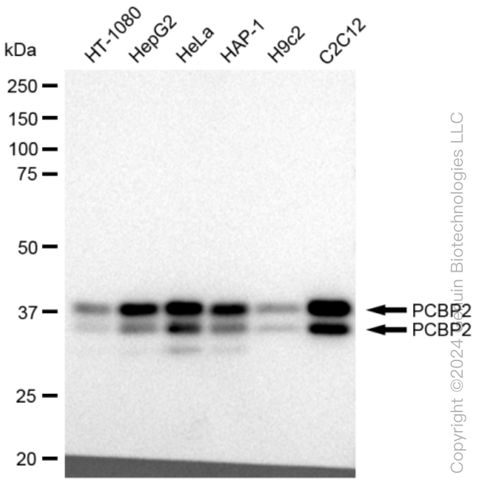
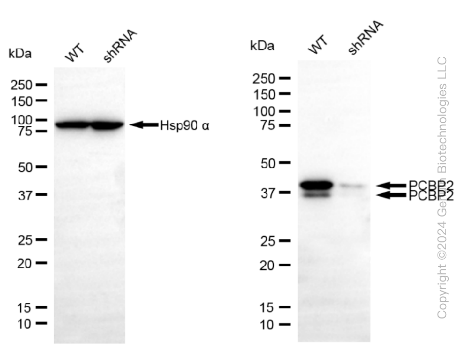
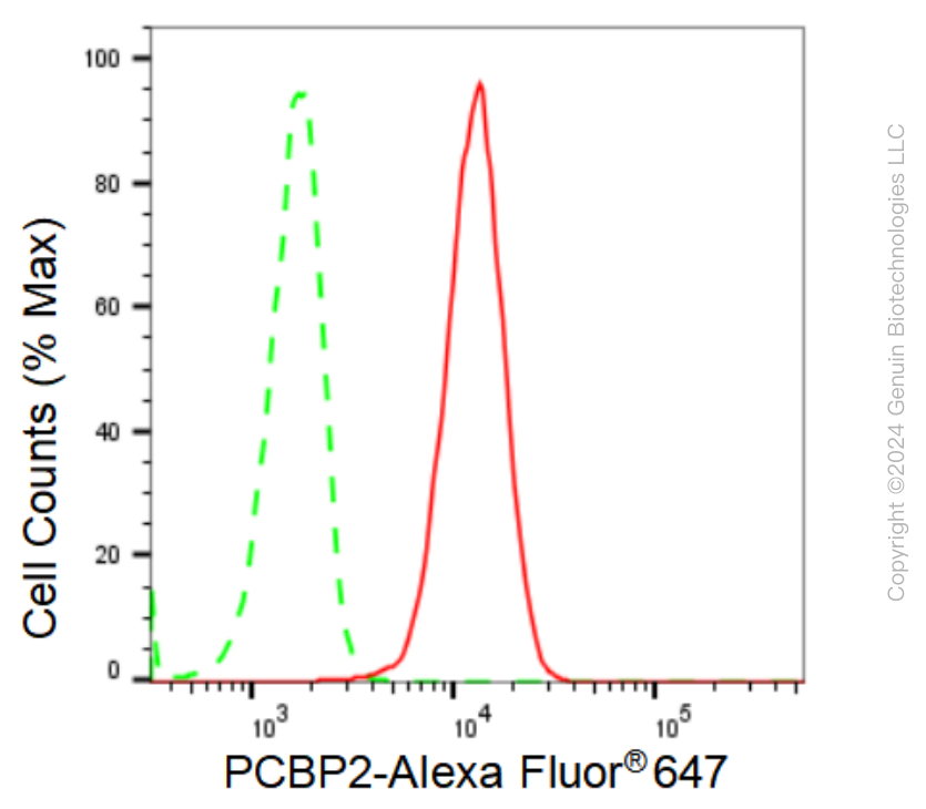
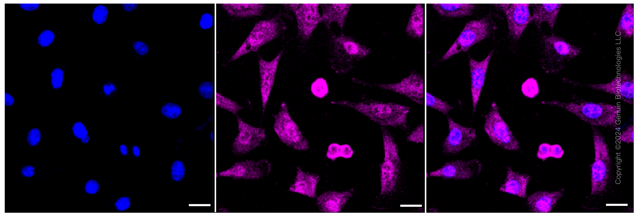
KD-Validated Anti-PCBP2 Rabbit Monoclonal Antibody
Rabbit monoclonal antibody
- SPECIFICATION
- CITATIONS
- PROTOCOLS
- BACKGROUND

Application
| WB, FC, ICC |
|---|---|
| Primary Accession | Q15366 |
| Reactivity | Rat, Human, Mouse |
| Clonality | Monoclonal |
| Isotype | Rabbit IgG |
| Clone Names | 23GB 2110 |
| Calculated MW | Predicted, 34-39 kDa , observed, 35,39 kDa |
| Gene Name | PCBP2 |
| Aliases | PCBP2; Poly(RC) Binding Protein 2; Poly(RC)-Binding Protein 2; HNRNPE2; HNRPE2; Heterogeneous Nuclear Ribonucleoprotein E2; Heterogenous Nuclear Ribonucleoprotein E2; Alpha-CP2; HnRNP-E2; HnRNP E2; Epididymis Secretory Sperm Binding Protein; HNRNP-E2 |
| Immunogen | A synthesized peptide derived from human PCBP2 |
| Gene ID | 5094 |
|---|---|
| Other Names | Poly(rC)-binding protein 2, Alpha-CP2, Heterogeneous nuclear ribonucleoprotein E2, hnRNP E2, PCBP2 {ECO:0000303|PubMed:7607214, ECO:0000312|HGNC:HGNC:8648} |
| Name | PCBP2 {ECO:0000303|PubMed:7607214, ECO:0000312|HGNC:HGNC:8648} |
|---|---|
| Function | Single-stranded nucleic acid binding protein that binds preferentially to oligo dC (PubMed:12414943, PubMed:7607214). Major cellular poly(rC)-binding protein (PubMed:12414943). Also binds poly(rU) (PubMed:12414943). Acts as a negative regulator of antiviral signaling (PubMed:19881509, PubMed:35322803). Negatively regulates cellular antiviral responses mediated by MAVS signaling (PubMed:19881509). It acts as an adapter between MAVS and the E3 ubiquitin ligase ITCH, therefore triggering MAVS ubiquitination and degradation (PubMed:19881509). Negativeley regulates the cGAS-STING pathway via interaction with CGAS, preventing the formation of liquid- like droplets in which CGAS is activated (PubMed:35322803). Together with PCBP1, required for erythropoiesis, possibly by regulating mRNA splicing (By similarity). |
| Cellular Location | Nucleus. Cytoplasm. Note=Loosely bound in the nucleus (PubMed:7607214). May shuttle between the nucleus and the cytoplasm (PubMed:7607214). |
| Tissue Location | Detected in all tissues examined. |
Research Areas
Citations (0)

Thousands of laboratories across the world have published research that depended on the performance of antibodies from Abcepta to advance their research. Check out links to articles that cite our products in major peer-reviewed journals, organized by research category.
Submit your citation using an Abcepta antibody to
info@abcepta.com, and receive a free "I Love Antibodies" mug.
info@abcepta.com, and receive a free "I Love Antibodies" mug.

Application Protocols
Provided below are standard protocols that you may find useful for product applications.
Abcepta welcomes feedback from its customers.
If you have used an Abcepta product and would like to share how it has performed, please click on the "Submit Review" button and provide the requested information. Our staff will examine and post your review and contact you if needed.
If you have any additional inquiries please email technical services at tech@abcepta.com.
$ 399.20

$ 149.00
Cat# AGI1061
Ordering Information
United States
AlbaniaAustraliaAustriaBelgiumBosnia & HerzegovinaBrazilBulgariaCanadaCentral AmericaChinaCroatiaCyprusCzech RepublicDenmarkEstoniaFinlandFranceGermanyGreeceHong KongHungaryIcelandIndiaIndonesiaIrelandIsraelItalyJapanLatviaLithuaniaLuxembourgMacedoniaMalaysiaMaltaMexicoNetherlandsNew ZealandNorwayPakistanPolandPortugalRomaniaSerbiaSingaporeSlovakiaSloveniaSouth AfricaSouth KoreaSpainSwedenSwitzerlandTaiwanTurkeyUnited KingdomUnited StatesVietnamWorldwideOthers
USA Headquarters
(888) 735-7227 / (858) 622-0099 or (858) 875-1900
Other Products
Shipping Information
Domestic orders (in stock items)
Shipped out the same day. Orders placed after 1 PM (PST) will ship out the next business day.
International orders
Contact your local distributors
