


 Foundational characteristics of cancer include proliferation, angiogenesis, migration, evasion of apoptosis, and cellular immortality. Find key markers for these cellular processes and antibodies to detect them.
 The SUMOplot™ Analysis Program predicts and scores sumoylation sites in your protein. SUMOylation is a post-translational modification involved in various cellular processes, such as nuclear-cytosolic transport, transcriptional regulation, apoptosis, protein stability, response to stress, and progression through the cell cycle.
 The Autophagy Receptor Motif Plotter predicts and scores autophagy receptor binding sites in your protein. Identifying proteins connected to this pathway is critical to understanding the role of autophagy in physiological as well as pathological processes such as development, differentiation, neurodegenerative diseases, stress, infection, and cancer.
> home > Products > Primary Antibodies > Antibody Collections > KD-Validated Antibodies > KD-Validated Anti-CLIP1 Rabbit Monoclonal Antibody
KD-Validated Anti-CLIP1 Rabbit Monoclonal Antibody
Rabbit monoclonal antibody
- SPECIFICATION
- CITATIONS
- PROTOCOLS
- BACKGROUND

Application
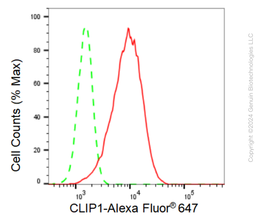
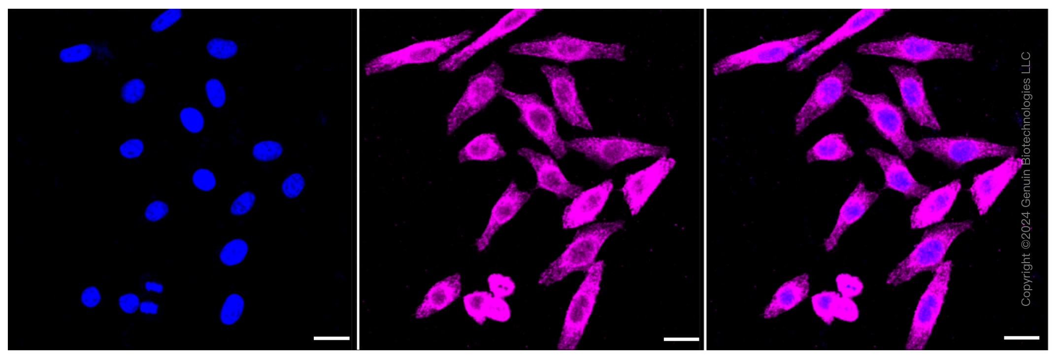
| WB, FC, ICC |
|---|---|
| Primary Accession | P30622 |
| Reactivity | Human, Mouse |
| Clonality | Monoclonal |
| Isotype | Rabbit IgG |
| Clone Names | 23GB3080 |
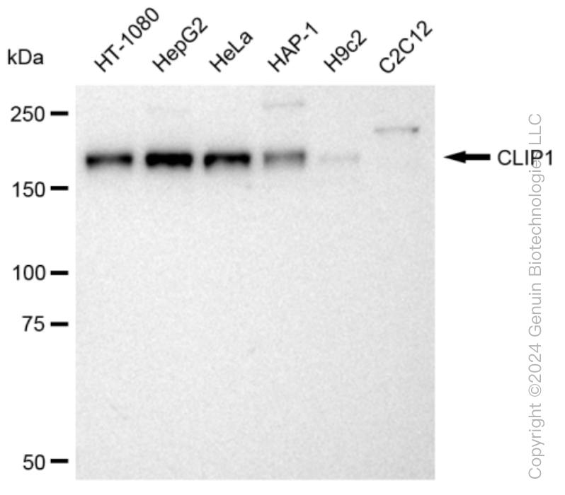
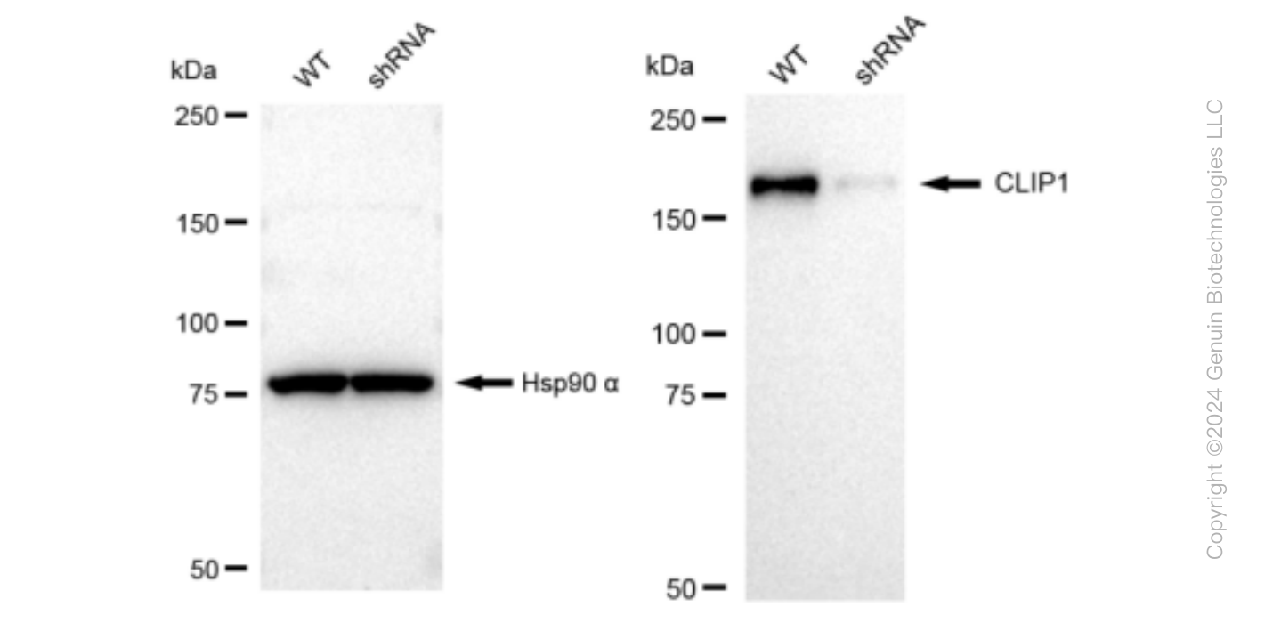
| Calculated MW | Predicted, 162 kDa , observed, 170 kDa |
| Gene Name | CLIP1 |
| Aliases | CLIP1; CAP-Gly Domain Containing Linker Protein 1; CLIP-170; CYLN1; CLIP170; CLIP; RSN; Restin (Reed-Steinberg Cell-Expressed Intermediate Filament-Associated Protein); CAP-Gly Domain-Containing Linker Protein 1; Cytoplasmic Linker Protein 170 Alpha-2; Cytoplasmic Linker Protein 1; Restin; Reed-Sternberg Intermediate Filament-Associated Protein; Cytoplasmic Linker Protein CLIP-170 |
| Immunogen | A synthesized peptide derived from CLIP170 |
| Gene ID | 6249 |
|---|---|
| Other Names | CAP-Gly domain-containing linker protein 1, Cytoplasmic linker protein 1, Cytoplasmic linker protein 170 alpha-2, CLIP-170, Reed-Sternberg intermediate filament-associated protein, Restin, CLIP1, CYLN1, RSN |
| Name | CLIP1 |
|---|---|
| Synonyms | CYLN1, RSN |
| Function | Binds to the plus end of microtubules and regulates the dynamics of the microtubule cytoskeleton. Promotes microtubule growth and microtubule bundling. Links cytoplasmic vesicles to microtubules and thereby plays an important role in intracellular vesicle trafficking. Plays a role macropinocytosis and endosome trafficking. |
| Cellular Location | Cytoplasm. Cytoplasm, cytoskeleton. Cytoplasmic vesicle membrane; Peripheral membrane protein; Cytoplasmic side. Cell projection, ruffle. Note=Localizes to microtubule plus ends (PubMed:17889670, PubMed:21646404). Localizes preferentially to the ends of tyrosinated microtubules (By similarity). Accumulates in plasma membrane regions with ruffling and protrusions. Associates with the membranes of intermediate macropinocytic vesicles (PubMed:12433698) {ECO:0000250|UniProtKB:Q922J3, ECO:0000269|PubMed:12433698, ECO:0000269|PubMed:17889670, ECO:0000269|PubMed:21646404} |
| Tissue Location | Detected in dendritic cells (at protein level). Highly expressed in the Reed-Sternberg cells of Hodgkin disease |
Research Areas
Citations (0)

Thousands of laboratories across the world have published research that depended on the performance of antibodies from Abcepta to advance their research. Check out links to articles that cite our products in major peer-reviewed journals, organized by research category.
Submit your citation using an Abcepta antibody to
info@abcepta.com, and receive a free "I Love Antibodies" mug.
info@abcepta.com, and receive a free "I Love Antibodies" mug.

Application Protocols
Provided below are standard protocols that you may find useful for product applications.
Abcepta welcomes feedback from its customers.
If you have used an Abcepta product and would like to share how it has performed, please click on the "Submit Review" button and provide the requested information. Our staff will examine and post your review and contact you if needed.
If you have any additional inquiries please email technical services at tech@abcepta.com.
$ 399.20

$ 149.00
Cat# AGI1157
Ordering Information
United States
AlbaniaAustraliaAustriaBelgiumBosnia & HerzegovinaBrazilBulgariaCanadaCentral AmericaChinaCroatiaCyprusCzech RepublicDenmarkEstoniaFinlandFranceGermanyGreeceHong KongHungaryIcelandIndiaIndonesiaIrelandIsraelItalyJapanLatviaLithuaniaLuxembourgMacedoniaMalaysiaMaltaMexicoNetherlandsNew ZealandNorwayPakistanPolandPortugalRomaniaSerbiaSingaporeSlovakiaSloveniaSouth AfricaSouth KoreaSpainSwedenSwitzerlandTaiwanTurkeyUnited KingdomUnited StatesVietnamWorldwideOthers
USA Headquarters
(888) 735-7227 / (858) 622-0099 or (858) 875-1900
Other Products
Shipping Information
Domestic orders (in stock items)
Shipped out the same day. Orders placed after 1 PM (PST) will ship out the next business day.
International orders
Contact your local distributors
