


 Foundational characteristics of cancer include proliferation, angiogenesis, migration, evasion of apoptosis, and cellular immortality. Find key markers for these cellular processes and antibodies to detect them.
 The SUMOplot™ Analysis Program predicts and scores sumoylation sites in your protein. SUMOylation is a post-translational modification involved in various cellular processes, such as nuclear-cytosolic transport, transcriptional regulation, apoptosis, protein stability, response to stress, and progression through the cell cycle.
 The Autophagy Receptor Motif Plotter predicts and scores autophagy receptor binding sites in your protein. Identifying proteins connected to this pathway is critical to understanding the role of autophagy in physiological as well as pathological processes such as development, differentiation, neurodegenerative diseases, stress, infection, and cancer.
> home > Products > Primary Antibodies > Antibody Collections > KD-Validated Antibodies > KD-Validated Anti-ADP ribosylation factor 6 Rabbit Monoclonal Antibody
KD-Validated Anti-ADP ribosylation factor 6 Rabbit Monoclonal Antibody
Rabbit monoclonal antibody
- SPECIFICATION
- CITATIONS
- PROTOCOLS
- BACKGROUND

Application
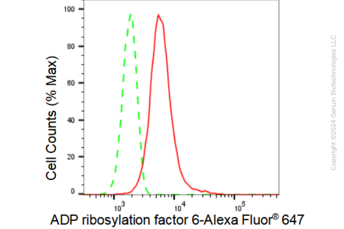
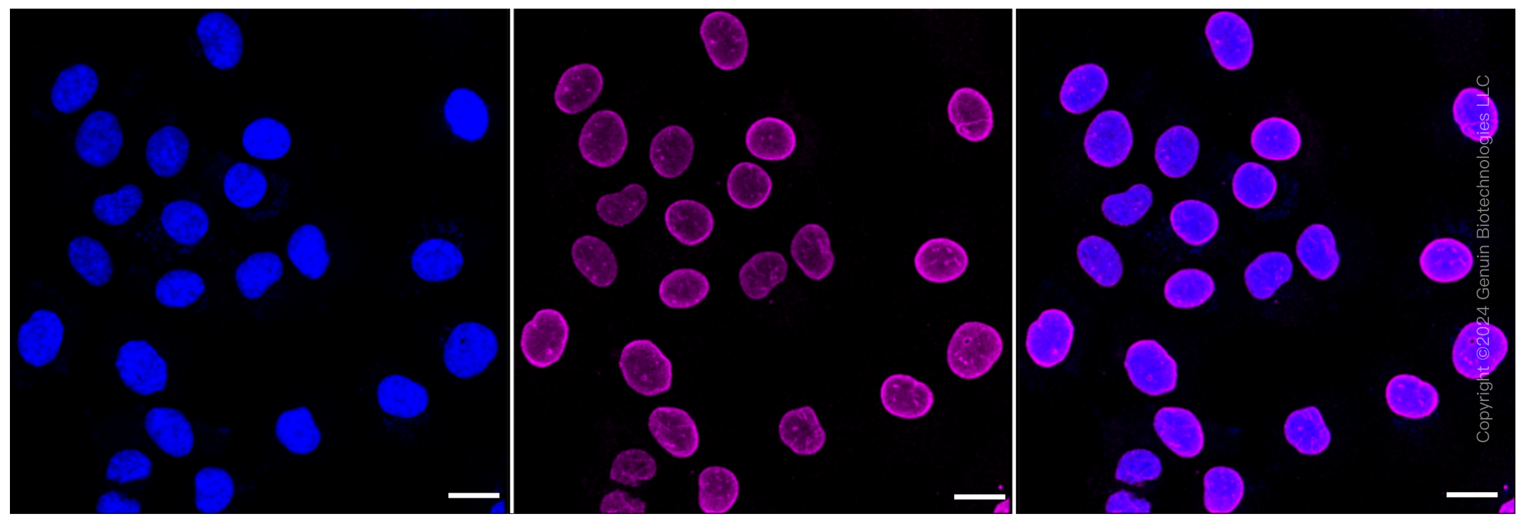
| WB, FC, ICC |
|---|---|
| Primary Accession | P62330 |
| Reactivity | Rat, Human, Mouse |
| Clonality | Monoclonal |
| Isotype | Rabbit IgG |
| Clone Names | 23GB3160 |
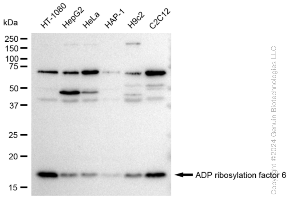
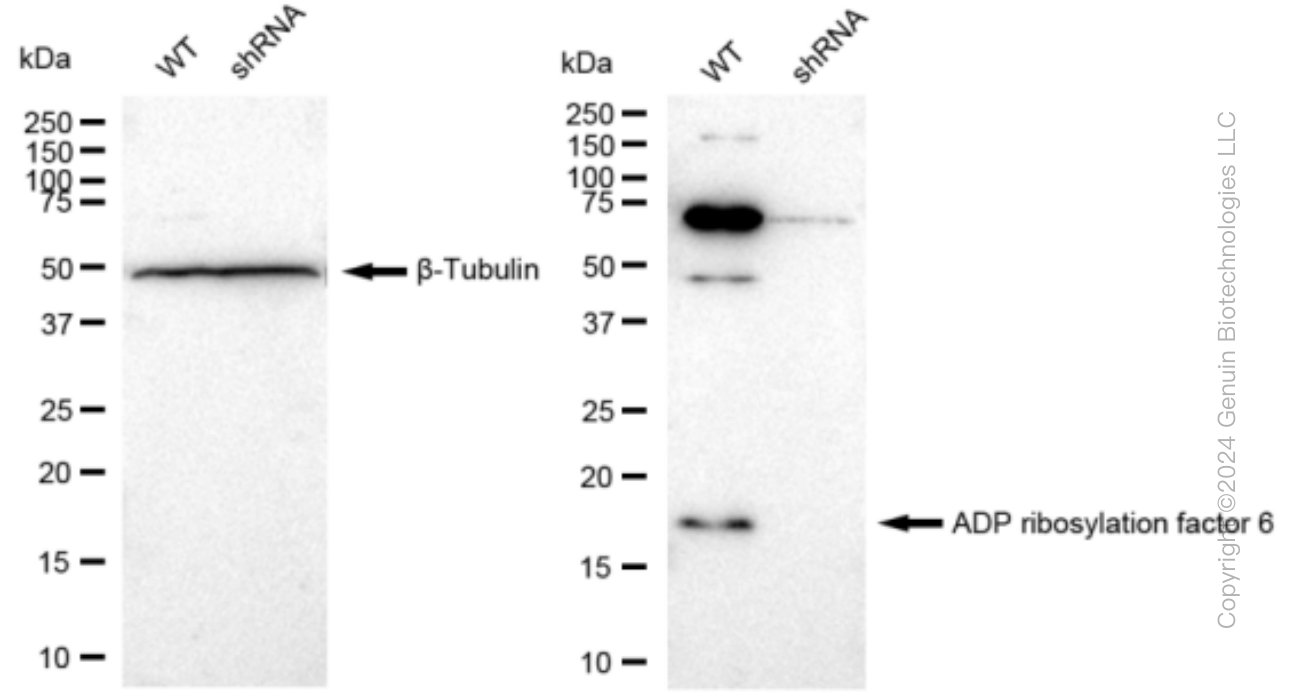
| Calculated MW | Predicted, 20 kDa, observed, 18 kDa |
| Gene Name | ARF6 |
| Aliases | ARF6; ADP Ribosylation Factor 6; ADP-Ribosylation Factor 6; EC 3.6.5.2 |
| Immunogen | A synthesized peptide derived from human ARF6 |
| Gene ID | 382 |
|---|---|
| Other Names | ADP-ribosylation factor 6 {ECO:0000303|Ref.6}, 3.6.5.2, ARF6 {ECO:0000303|Ref.6, ECO:0000312|HGNC:HGNC:659} |
| Name | ARF6 {ECO:0000303|Ref.6, ECO:0000312|HGNC:HGNC:659} |
|---|---|
| Function | GTP-binding protein involved in protein trafficking that regulates endocytic recycling and cytoskeleton remodeling (PubMed:11266366, PubMed:16737952, PubMed:18400762, PubMed:21170023, PubMed:32103017, PubMed:7589240). GTP-bound form plays an important role in the transport of multiple palmitoylated proteins form the Golgi to the plasma membrane (PubMed:37461827). Required for normal completion of mitotic cytokinesis (By similarity). Plays a role in the reorganization of the actin cytoskeleton and the formation of stress fibers (By similarity). Involved in the regulation of dendritic spine development, contributing to the regulation of dendritic branching and filopodia extension (PubMed:14978216). Potentiates the neurite outgrowth in primary neurons by interacting with the molecular adapter APBB1 (PubMed:36250347). Plays an important role in membrane trafficking, during junctional remodeling and epithelial polarization (PubMed:36017701). Regulates surface levels of adherens junction proteins such as CDH1 (By similarity). Required for NTRK1 sorting to the recycling pathway from early endosomes (By similarity). |
| Cellular Location | Cytoplasm, cytosol. Cell membrane; Lipid-anchor. Endosome membrane; Lipid-anchor. Recycling endosome membrane; Lipid-anchor. Cell projection, filopodium membrane; Lipid- anchor. Cell projection, ruffle. Cleavage furrow. Midbody, Midbody ring. Early endosome membrane {ECO:0000250|UniProtKB:P62331}; Lipid-anchor {ECO:0000250|UniProtKB:P62331}. Golgi apparatus, trans-Golgi network membrane {ECO:0000250|UniProtKB:P62331}; Lipid-anchor {ECO:0000250|UniProtKB:P62331}. Note=Distributed uniformly on the plasma membrane, as well as throughout the cytoplasm during metaphase Subsequently concentrated at patches in the equatorial region at the onset of cytokinesis, and becomes distributed in the equatorial region concurrent with cleavage furrow ingression. In late stages of cytokinesis, concentrates at the midbody ring/Flemming body (PubMed:23603394). Recruitment to the midbody ring requires both activation by PSD/EFA6A and interaction with KIF23/MKLP1 (PubMed:23603394). After abscission of the intercellular bridge, incorporated into one of the daughter cells as a midbody remnant and localizes to punctate structures beneath the plasma membrane (PubMed:23603394). Recruited to the cell membrane in association with CYTH2 and ARL4C (PubMed:17398095). Colocalizes with DAB2IP at the plasma membrane and endocytic vesicles (PubMed:19948740) Myristoylation is required for proper localization to membranes: myristoylation on Lys-3 allows ARF6 to remain on membranes during the GTPase cycle (PubMed:32103017, PubMed:7589240) |
| Tissue Location | Ubiquitous, with higher levels in heart, substantia nigra, and kidney. |
Research Areas
Citations (0)

Thousands of laboratories across the world have published research that depended on the performance of antibodies from Abcepta to advance their research. Check out links to articles that cite our products in major peer-reviewed journals, organized by research category.
Submit your citation using an Abcepta antibody to
info@abcepta.com, and receive a free "I Love Antibodies" mug.
info@abcepta.com, and receive a free "I Love Antibodies" mug.

Application Protocols
Provided below are standard protocols that you may find useful for product applications.
Abcepta welcomes feedback from its customers.
If you have used an Abcepta product and would like to share how it has performed, please click on the "Submit Review" button and provide the requested information. Our staff will examine and post your review and contact you if needed.
If you have any additional inquiries please email technical services at tech@abcepta.com.
$ 149.00
$ 499.00
Cat# AGI1190
Ordering Information
United States
AlbaniaAustraliaAustriaBelgiumBosnia & HerzegovinaBrazilBulgariaCanadaCentral AmericaChinaCroatiaCyprusCzech RepublicDenmarkEstoniaFinlandFranceGermanyGreeceHong KongHungaryIcelandIndiaIndonesiaIrelandIsraelItalyJapanLatviaLithuaniaLuxembourgMacedoniaMalaysiaMaltaMexicoNetherlandsNew ZealandNorwayPakistanPolandPortugalRomaniaSerbiaSingaporeSlovakiaSloveniaSouth AfricaSouth KoreaSpainSwedenSwitzerlandTaiwanTurkeyUnited KingdomUnited StatesVietnamWorldwideOthers
USA Headquarters
(888) 735-7227 / (858) 622-0099 or (858) 875-1900
Other Products
Shipping Information
Domestic orders (in stock items)
Shipped out the same day. Orders placed after 1 PM (PST) will ship out the next business day.
International orders
Contact your local distributors
