


 Foundational characteristics of cancer include proliferation, angiogenesis, migration, evasion of apoptosis, and cellular immortality. Find key markers for these cellular processes and antibodies to detect them.
 The SUMOplot™ Analysis Program predicts and scores sumoylation sites in your protein. SUMOylation is a post-translational modification involved in various cellular processes, such as nuclear-cytosolic transport, transcriptional regulation, apoptosis, protein stability, response to stress, and progression through the cell cycle.
 The Autophagy Receptor Motif Plotter predicts and scores autophagy receptor binding sites in your protein. Identifying proteins connected to this pathway is critical to understanding the role of autophagy in physiological as well as pathological processes such as development, differentiation, neurodegenerative diseases, stress, infection, and cancer.
> home > Products > Primary Antibodies > Antibody Collections > KD-Validated Antibodies > KD-Validated Anti-Dihydrolipoamide dehydrogenase Rabbit Monoclonal Antibody
KD-Validated Anti-Dihydrolipoamide dehydrogenase Rabbit Monoclonal Antibody
Rabbit monoclonal antibody
- SPECIFICATION
- CITATIONS
- PROTOCOLS
- BACKGROUND

Application
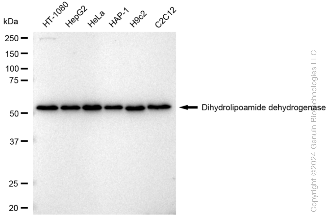
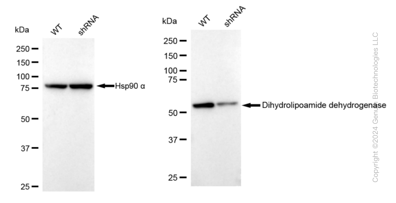
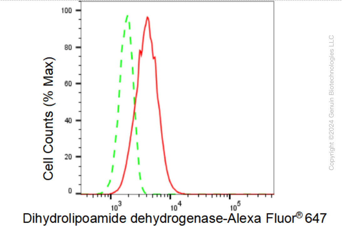
| WB, FC |
|---|---|
| Primary Accession | P09622 |
| Reactivity | Rat, Human, Mouse |
| Clonality | Monoclonal |
| Isotype | Rabbit IgG |
| Clone Names | 23GB3825 |
| Calculated MW | Predicted, 54 kDa , observed, 56 kDa |
| Gene Name | DLD |
| Aliases | DLD; Dihydrolipoamide Dehydrogenase; OGDC-E3; DLDH; GCSL; LAD; E3; E3 Component Of Pyruvate Dehydrogenase Complex, 2-Oxo-Glutarate Complex, Branched Chain Keto Acid Dehydrogenase Complex; Dihydrolipoyl Dehydrogenase, Mitochondrial; Glycine Cleavage System L Protein; EC 1.8.1.4; PHE3; Dihydrolipoamide Dehydrogenase (E3 Component Of Pyruvate Dehydrogenase Complex, 2-Oxo-Glutarate Complex, Branched Chain Keto Acid Dehydrogenase Complex); Epididymis Secretory Sperm Binding Protein; Glycine Cleavage System Protein L; Lipoamide Dehydrogenase; Lipoyl Dehydrogenase; Lipoamide Reductase; Diaphorase; EC 1.8.1 48; DLDD |
| Immunogen | A synthesized peptide derived from human DLDH |
| Gene ID | 1738 |
|---|---|
| Other Names | Dihydrolipoyl dehydrogenase, mitochondrial, 1.8.1.4, Dihydrolipoamide dehydrogenase, Glycine cleavage system L protein, DLD, GCSL, LAD, PHE3 |
| Name | DLD |
|---|---|
| Synonyms | GCSL, LAD, PHE3 |
| Function | Lipoamide dehydrogenase is a component of the glycine cleavage system as well as an E3 component of three alpha-ketoacid dehydrogenase complexes (pyruvate-, alpha-ketoglutarate-, and branched- chain amino acid-dehydrogenase complex) (PubMed:15712224, PubMed:16442803, PubMed:16770810, PubMed:17404228, PubMed:20160912, PubMed:20385101). The 2-oxoglutarate dehydrogenase complex is mainly active in the mitochondrion (PubMed:29211711). A fraction of the 2- oxoglutarate dehydrogenase complex also localizes in the nucleus and is required for lysine succinylation of histones: associates with KAT2A on chromatin and provides succinyl-CoA to histone succinyltransferase KAT2A (PubMed:29211711). In monomeric form may have additional moonlighting function as serine protease (PubMed:17404228). Involved in the hyperactivation of spermatazoa during capacitation and in the spermatazoal acrosome reaction (By similarity). |
| Cellular Location | Mitochondrion matrix. Nucleus. Cell projection, cilium, flagellum {ECO:0000250|UniProtKB:Q811C4}. Cytoplasmic vesicle, secretory vesicle, acrosome. Note=Mainly localizes in the mitochondrion. A small fraction localizes to the nucleus, where the 2- oxoglutarate dehydrogenase complex is required for histone succinylation. |
Research Areas
Citations (0)

Thousands of laboratories across the world have published research that depended on the performance of antibodies from Abcepta to advance their research. Check out links to articles that cite our products in major peer-reviewed journals, organized by research category.
Submit your citation using an Abcepta antibody to
info@abcepta.com, and receive a free "I Love Antibodies" mug.
info@abcepta.com, and receive a free "I Love Antibodies" mug.

Application Protocols
Provided below are standard protocols that you may find useful for product applications.
Abcepta welcomes feedback from its customers.
If you have used an Abcepta product and would like to share how it has performed, please click on the "Submit Review" button and provide the requested information. Our staff will examine and post your review and contact you if needed.
If you have any additional inquiries please email technical services at tech@abcepta.com.
$ 399.20

$ 149.00
Cat# AGI1232
Ordering Information
United States
AlbaniaAustraliaAustriaBelgiumBosnia & HerzegovinaBrazilBulgariaCanadaCentral AmericaChinaCroatiaCyprusCzech RepublicDenmarkEstoniaFinlandFranceGermanyGreeceHong KongHungaryIcelandIndiaIndonesiaIrelandIsraelItalyJapanLatviaLithuaniaLuxembourgMacedoniaMalaysiaMaltaMexicoNetherlandsNew ZealandNorwayPakistanPolandPortugalRomaniaSerbiaSingaporeSlovakiaSloveniaSouth AfricaSouth KoreaSpainSwedenSwitzerlandTaiwanTurkeyUnited KingdomUnited StatesVietnamWorldwideOthers
USA Headquarters
(888) 735-7227 / (858) 622-0099 or (858) 875-1900
Other Products
Shipping Information
Domestic orders (in stock items)
Shipped out the same day. Orders placed after 1 PM (PST) will ship out the next business day.
International orders
Contact your local distributors
