


 Foundational characteristics of cancer include proliferation, angiogenesis, migration, evasion of apoptosis, and cellular immortality. Find key markers for these cellular processes and antibodies to detect them.
 The SUMOplot™ Analysis Program predicts and scores sumoylation sites in your protein. SUMOylation is a post-translational modification involved in various cellular processes, such as nuclear-cytosolic transport, transcriptional regulation, apoptosis, protein stability, response to stress, and progression through the cell cycle.
 The Autophagy Receptor Motif Plotter predicts and scores autophagy receptor binding sites in your protein. Identifying proteins connected to this pathway is critical to understanding the role of autophagy in physiological as well as pathological processes such as development, differentiation, neurodegenerative diseases, stress, infection, and cancer.
> home > Products > Primary Antibodies > Antibody Collections > KD-Validated Antibodies > KD-Validated Anti-Phospholipase A2 activating protein Rabbit Monoclonal Antibody
KD-Validated Anti-Phospholipase A2 activating protein Rabbit Monoclonal Antibody
Rabbit monoclonal antibody
- SPECIFICATION
- CITATIONS
- PROTOCOLS
- BACKGROUND

Application
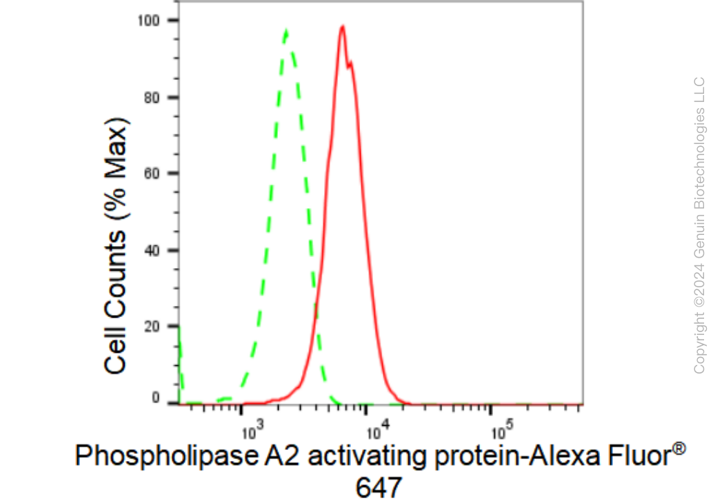
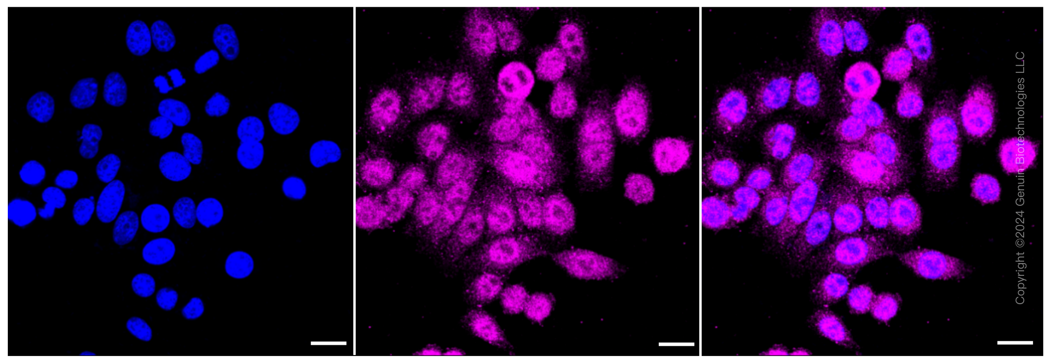
| WB, FC, ICC |
|---|---|
| Primary Accession | Q9Y263 |
| Reactivity | Human |
| Clonality | Monoclonal |
| Isotype | Rabbit IgG |
| Clone Names | 23GB 2560 |
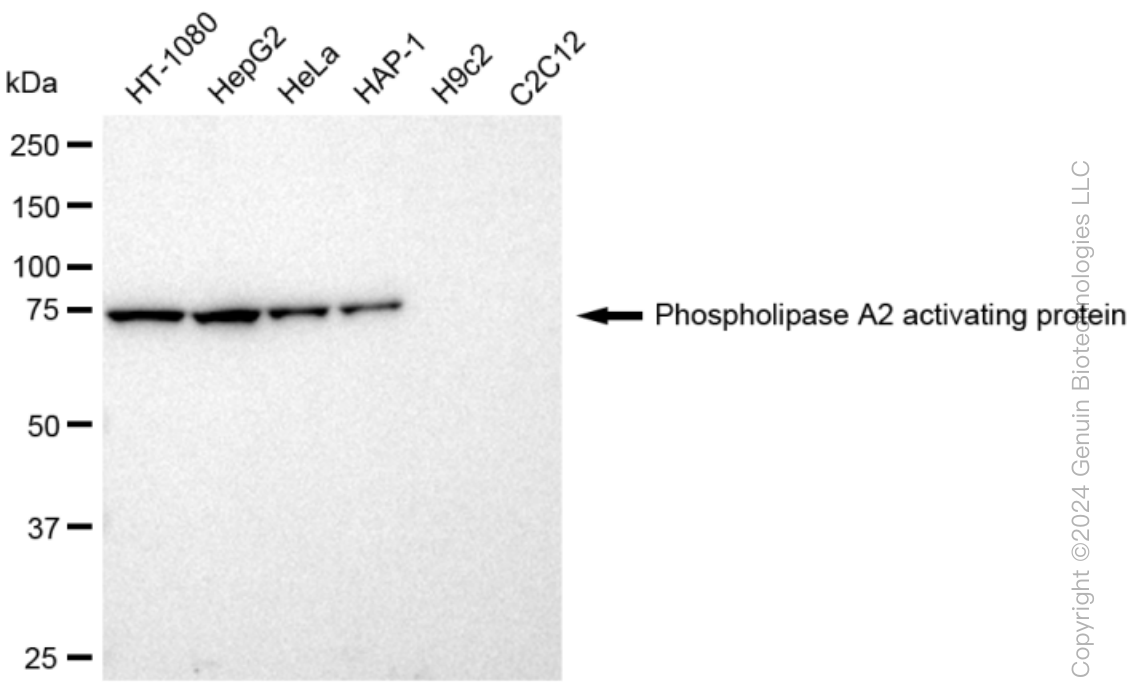
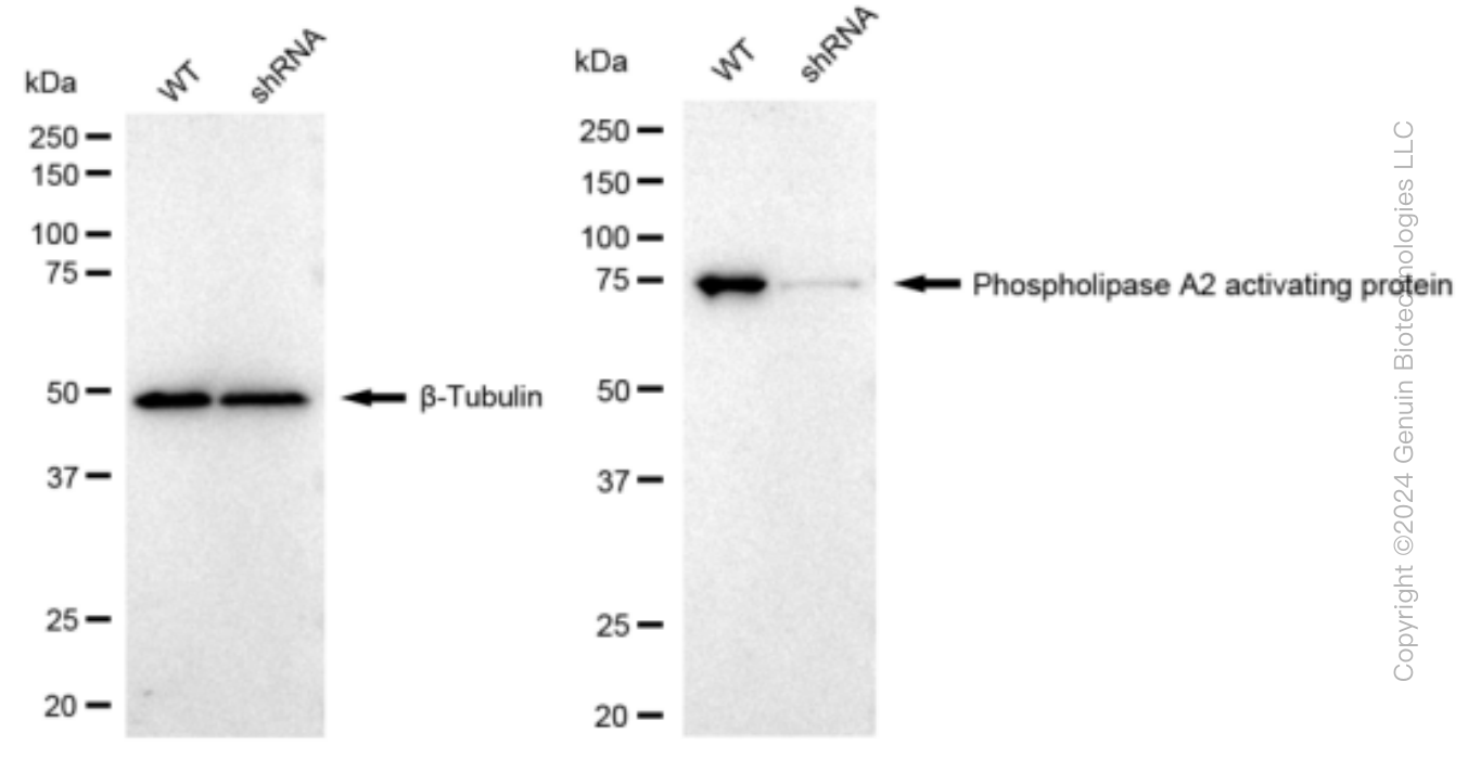
| Calculated MW | Predicted, 87 kDa , observed, 75 kDa |
| Gene Name | PLAA |
| Aliases | PLAA; Phospholipase A2 Activating Protein; PLA2P; PLAP; DOA1; Phospholipase A-2-Activating Protein; FLJ11281; FLJ12699; DOA1 Homolog (S. Cerevisiae); DOA1 Homolog; NDMSBA |
| Immunogen | A synthesized peptide derived from human PLAP |
| Gene ID | 9373 |
|---|---|
| Other Names | Phospholipase A-2-activating protein, PLA2P, PLAP, PLAA, PLAP |
| Name | PLAA |
|---|---|
| Synonyms | PLAP |
| Function | Plays a role in protein ubiquitination, sorting and degradation through its association with VCP (PubMed:27753622). Involved in ubiquitin-mediated membrane proteins trafficking to late endosomes in an ESCRT-dependent manner, and hence plays a role in synaptic vesicle recycling (By similarity). May play a role in macroautophagy, regulating for instance the clearance of damaged lysosomes (PubMed:27753622). Plays a role in cerebellar Purkinje cell development (By similarity). Positively regulates cytosolic and calcium-independent phospholipase A2 activities in a tumor necrosis factor alpha (TNF-alpha)- or lipopolysaccharide (LPS)-dependent manner, and hence prostaglandin E2 biosynthesis (PubMed:18291623, PubMed:28007986). |
| Cellular Location | Nucleus. Cytoplasm. Synapse {ECO:0000250|UniProtKB:P27612}. Note=Recruited to damaged lysosomes decorated with K48-linked ubiquitin chains |
Research Areas
Citations (0)

Thousands of laboratories across the world have published research that depended on the performance of antibodies from Abcepta to advance their research. Check out links to articles that cite our products in major peer-reviewed journals, organized by research category.
Submit your citation using an Abcepta antibody to
info@abcepta.com, and receive a free "I Love Antibodies" mug.
info@abcepta.com, and receive a free "I Love Antibodies" mug.

Application Protocols
Provided below are standard protocols that you may find useful for product applications.
Abcepta welcomes feedback from its customers.
If you have used an Abcepta product and would like to share how it has performed, please click on the "Submit Review" button and provide the requested information. Our staff will examine and post your review and contact you if needed.
If you have any additional inquiries please email technical services at tech@abcepta.com.
$ 399.20

$ 149.00
Cat# AGI1267
Ordering Information
United States
AlbaniaAustraliaAustriaBelgiumBosnia & HerzegovinaBrazilBulgariaCanadaCentral AmericaChinaCroatiaCyprusCzech RepublicDenmarkEstoniaFinlandFranceGermanyGreeceHong KongHungaryIcelandIndiaIndonesiaIrelandIsraelItalyJapanLatviaLithuaniaLuxembourgMacedoniaMalaysiaMaltaMexicoNetherlandsNew ZealandNorwayPakistanPolandPortugalRomaniaSerbiaSingaporeSlovakiaSloveniaSouth AfricaSouth KoreaSpainSwedenSwitzerlandTaiwanTurkeyUnited KingdomUnited StatesVietnamWorldwideOthers
USA Headquarters
(888) 735-7227 / (858) 622-0099 or (858) 875-1900
Other Products
Shipping Information
Domestic orders (in stock items)
Shipped out the same day. Orders placed after 1 PM (PST) will ship out the next business day.
International orders
Contact your local distributors
