


 Foundational characteristics of cancer include proliferation, angiogenesis, migration, evasion of apoptosis, and cellular immortality. Find key markers for these cellular processes and antibodies to detect them.
 The SUMOplot™ Analysis Program predicts and scores sumoylation sites in your protein. SUMOylation is a post-translational modification involved in various cellular processes, such as nuclear-cytosolic transport, transcriptional regulation, apoptosis, protein stability, response to stress, and progression through the cell cycle.
 The Autophagy Receptor Motif Plotter predicts and scores autophagy receptor binding sites in your protein. Identifying proteins connected to this pathway is critical to understanding the role of autophagy in physiological as well as pathological processes such as development, differentiation, neurodegenerative diseases, stress, infection, and cancer.
> home > Products > Primary Antibodies > Antibody Collections > KD-Validated Antibodies > KD-Validated Anti-PTPN11 Rabbit Monoclonal Antibody
KD-Validated Anti-PTPN11 Rabbit Monoclonal Antibody
Rabbit monoclonal antibody
- SPECIFICATION
- CITATIONS
- PROTOCOLS
- BACKGROUND

Application
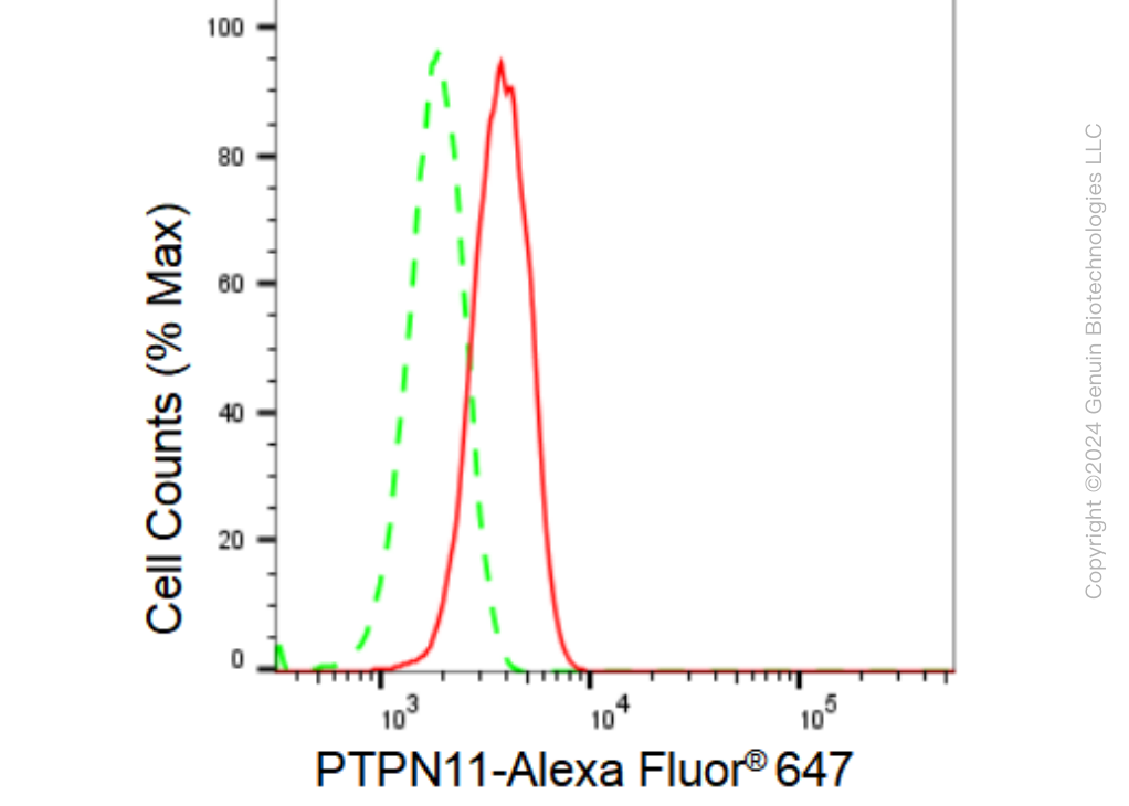
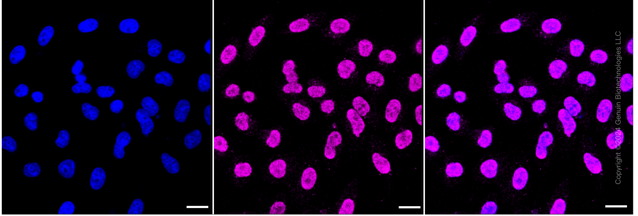
| WB, FC, ICC |
|---|---|
| Primary Accession | Q06124 |
| Reactivity | Rat, Human, Mouse |
| Clonality | Monoclonal |
| Isotype | Rabbit IgG |
| Clone Names | 23GB2670 |
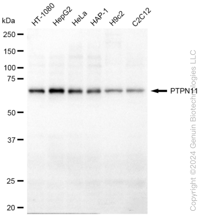
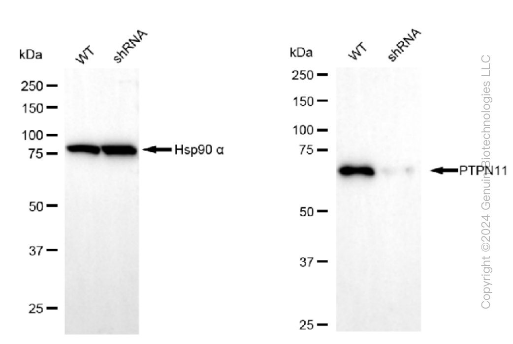
| Calculated MW | Predicted, 68 kDa , observed, 68 kDa |
| Gene Name | PTPN11 |
| Aliases | PTPN11; Protein Tyrosine Phosphatase Non-Receptor Type 11; SH-PTP2; PTP2C; BPTP3; SHP-2; SHP2; SH2 Domain-Containing Protein Tyrosine Phosphatase 2; Tyrosine-Protein Phosphatase Non-Receptor Type 11; Protein-Tyrosine Phosphatase 1D; Protein-Tyrosine Phosphatase 2C; EC 3.1.3.48; SH-PTP3; PTP-1D; NS1; Protein Tyrosine Phosphatase, Non-Receptor Type 11; Noonan Syndrome 1; METCDS; SHPTP2; PTP-2C; JMML; Shp2; CFC |
| Immunogen | A synthesized peptide derived from human SHP2 |
| Gene ID | 5781 |
|---|---|
| Other Names | Tyrosine-protein phosphatase non-receptor type 11, 3.1.3.48, Protein-tyrosine phosphatase 1D, PTP-1D, Protein-tyrosine phosphatase 2C, PTP-2C, SH-PTP2, SHP-2, Shp2, SH-PTP3, PTPN11, PTP2C, SHPTP2 |
| Name | PTPN11 |
|---|---|
| Synonyms | PTP2C, SHPTP2 |
| Function | Acts downstream of various receptor and cytoplasmic protein tyrosine kinases to participate in the signal transduction from the cell surface to the nucleus (PubMed:10655584, PubMed:14739280, PubMed:18559669, PubMed:18829466, PubMed:26742426, PubMed:28074573). Positively regulates MAPK signal transduction pathway (PubMed:28074573). Dephosphorylates GAB1, ARHGAP35 and EGFR (PubMed:28074573). Dephosphorylates ROCK2 at 'Tyr-722' resulting in stimulation of its RhoA binding activity (PubMed:18559669). Dephosphorylates CDC73 (PubMed:26742426). Dephosphorylates SOX9 on tyrosine residues, leading to inactivate SOX9 and promote ossification (By similarity). Dephosphorylates tyrosine-phosphorylated NEDD9/CAS-L (PubMed:19275884). |
| Cellular Location | Cytoplasm. Nucleus |
| Tissue Location | Widely expressed, with highest levels in heart, brain, and skeletal muscle. |
Research Areas
Citations (0)

Thousands of laboratories across the world have published research that depended on the performance of antibodies from Abcepta to advance their research. Check out links to articles that cite our products in major peer-reviewed journals, organized by research category.
Submit your citation using an Abcepta antibody to
info@abcepta.com, and receive a free "I Love Antibodies" mug.
info@abcepta.com, and receive a free "I Love Antibodies" mug.

Application Protocols
Provided below are standard protocols that you may find useful for product applications.
Abcepta welcomes feedback from its customers.
If you have used an Abcepta product and would like to share how it has performed, please click on the "Submit Review" button and provide the requested information. Our staff will examine and post your review and contact you if needed.
If you have any additional inquiries please email technical services at tech@abcepta.com.
$ 149.00
$ 499.00
Cat# AGI1274
Ordering Information
United States
AlbaniaAustraliaAustriaBelgiumBosnia & HerzegovinaBrazilBulgariaCanadaCentral AmericaChinaCroatiaCyprusCzech RepublicDenmarkEstoniaFinlandFranceGermanyGreeceHong KongHungaryIcelandIndiaIndonesiaIrelandIsraelItalyJapanLatviaLithuaniaLuxembourgMacedoniaMalaysiaMaltaMexicoNetherlandsNew ZealandNorwayPakistanPolandPortugalRomaniaSerbiaSingaporeSlovakiaSloveniaSouth AfricaSouth KoreaSpainSwedenSwitzerlandTaiwanTurkeyUnited KingdomUnited StatesVietnamWorldwideOthers
USA Headquarters
(888) 735-7227 / (858) 622-0099 or (858) 875-1900
Other Products
Shipping Information
Domestic orders (in stock items)
Shipped out the same day. Orders placed after 1 PM (PST) will ship out the next business day.
International orders
Contact your local distributors
