


 Foundational characteristics of cancer include proliferation, angiogenesis, migration, evasion of apoptosis, and cellular immortality. Find key markers for these cellular processes and antibodies to detect them.
 The SUMOplot™ Analysis Program predicts and scores sumoylation sites in your protein. SUMOylation is a post-translational modification involved in various cellular processes, such as nuclear-cytosolic transport, transcriptional regulation, apoptosis, protein stability, response to stress, and progression through the cell cycle.
 The Autophagy Receptor Motif Plotter predicts and scores autophagy receptor binding sites in your protein. Identifying proteins connected to this pathway is critical to understanding the role of autophagy in physiological as well as pathological processes such as development, differentiation, neurodegenerative diseases, stress, infection, and cancer.
> home > Products > Primary Antibodies > Antibody Collections > KD-Validated Antibodies > KD-Validated Anti-AMBP Rabbit Monoclonal Antibody
KD-Validated Anti-AMBP Rabbit Monoclonal Antibody
Rabbit monoclonal antibody
- SPECIFICATION
- CITATIONS
- PROTOCOLS
- BACKGROUND

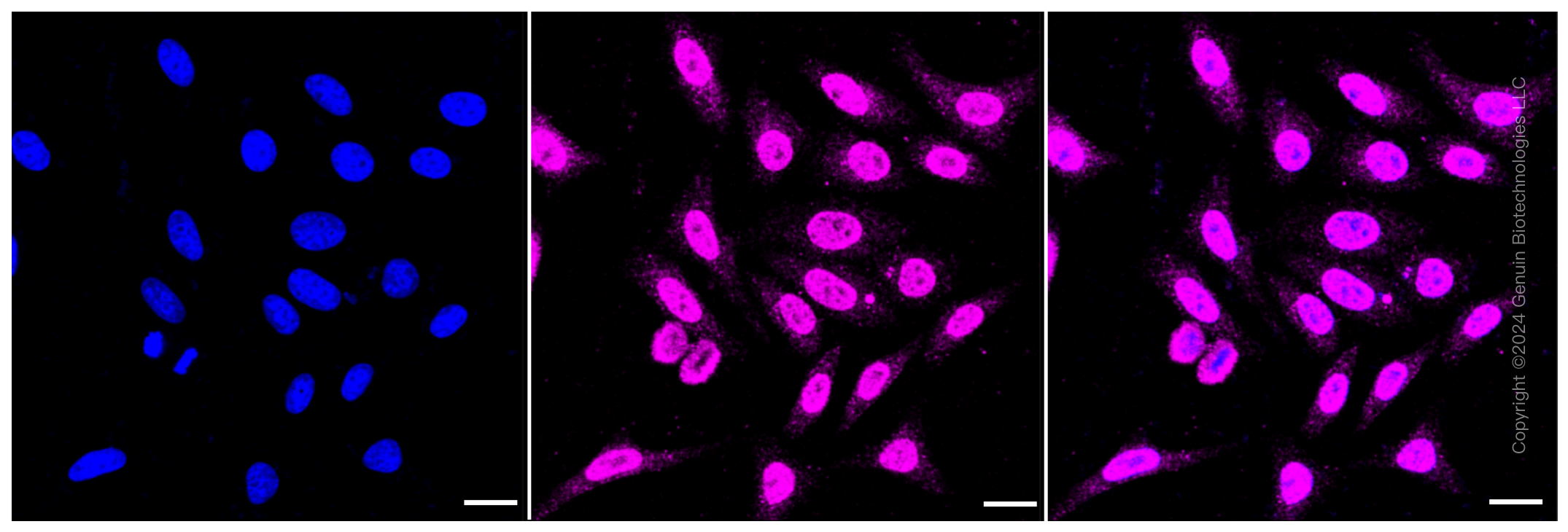
Application
| WB, ICC |
|---|---|
| Primary Accession | P02760 |
| Reactivity | Human |
| Clonality | Monoclonal |
| Isotype | Rabbit IgG |
| Clone Names | 23GB3200 |
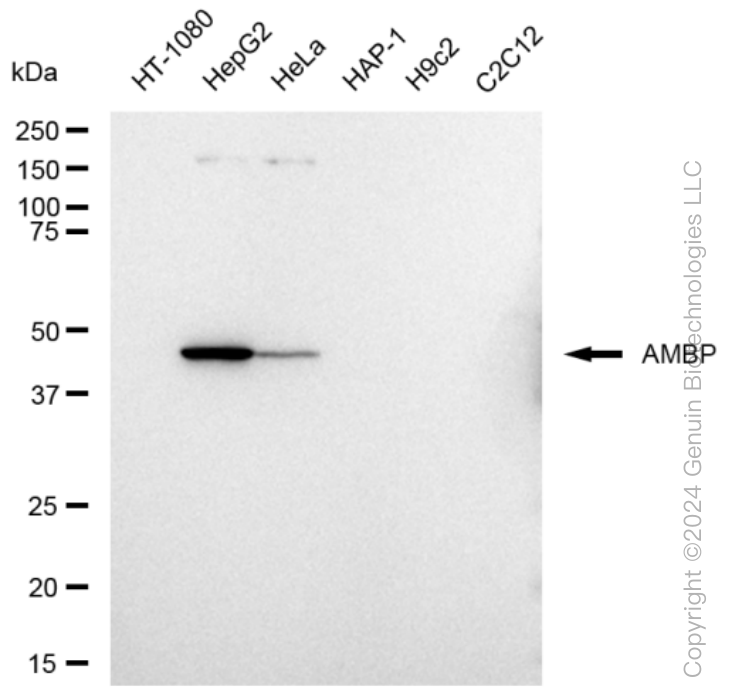
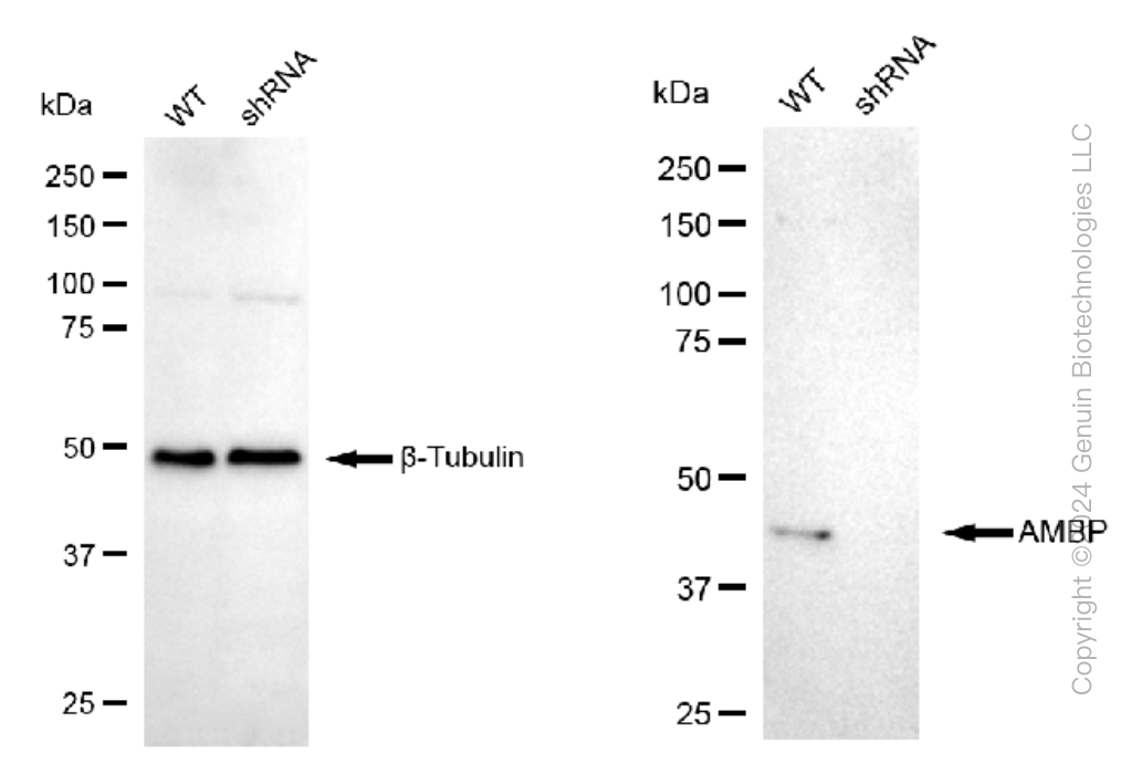
| Calculated MW | Predicted, 39 kDa , observed, 39 kDa |
| Gene Name | AMBP |
| Aliases | AMBP; Alpha-1-Microglobulin/Bikunin Precursor; HCP; Protein HC; IATIL; ITILC; EDC1; HI30; ITIL; UTI; Complex-Forming Glycoprotein Heterogeneous In Charge; Inter-Alpha-Trypsin Inhibitor Light Chain; Growth-Inhibiting Protein 19; Uronic-Acid-Rich Protein; Protein AMBP; Trypstatin; Uristatin; Bikunin; ITI; A1M |
| Immunogen | A synthesized peptide derived from human AMBP |
| Gene ID | 259 |
|---|---|
| Other Names | Protein AMBP, Protein HC, Alpha-1-microglobulin, 1.6.2.-, Alpha-1 microglycoprotein, Complex-forming glycoprotein heterogeneous in charge, Inter-alpha-trypsin inhibitor light chain, ITI-LC, Bikunin, EDC1, HI-30, Uronic-acid-rich protein, Trypstatin, AMBP, HCP, ITIL |
| Name | AMBP |
|---|---|
| Synonyms | HCP, ITIL |
| Function | [Alpha-1-microglobulin]: Antioxidant and tissue repair protein with reductase, heme-binding and radical-scavenging activities. Removes and protects against harmful oxidants and repairs macromolecules in intravascular and extravascular spaces and in intracellular compartments (PubMed:11877257, PubMed:15683711, PubMed:22096585, PubMed:23157686, PubMed:23642167, PubMed:25698971, PubMed:32092412, PubMed:32823731). Intravascularly, plays a regulatory role in red cell homeostasis by preventing heme- and reactive oxygen species-induced cell damage. Binds and degrades free heme to protect fetal and adult red blood cells from hemolysis (PubMed:11877257, PubMed:32092412). Reduces extracellular methemoglobin, a Fe3+ (ferric) form of hemoglobin that cannot bind oxygen, back to the Fe2+ (ferrous) form deoxyhemoglobin, which has oxygen-carrying potential (PubMed:15683711). Upon acute inflammation, inhibits oxidation of low- density lipoprotein particles by MPO and limits vascular damage (PubMed:25698971). Extravascularly, protects from oxidation products formed on extracellular matrix structures and cell membranes. Catalyzes the reduction of carbonyl groups on oxidized collagen fibers and preserves cellular and extracellular matrix ultrastructures (PubMed:22096585, PubMed:23642167). Importantly, counteracts the oxidative damage at blood-placenta interface, preventing leakage of free fetal hemoglobin into the maternal circulation (PubMed:21356557). Intracellularly, has a role in maintaining mitochondrial redox homeostasis. Bound to complex I of the respiratory chain of mitochondria, may scavenge free radicals and preserve mitochondrial ATP synthesis. Protects renal tubule epithelial cells from heme-induced oxidative damage to mitochondria (PubMed:23157686, PubMed:32823731). Reduces cytochrome c from Fe3+ (ferric) to the Fe2+ (ferrous) state through formation of superoxide anion radicals in the presence of ascorbate or NADH/NADPH electron donor cofactors, ascorbate being the preferred cofactor (PubMed:15683711). Has a chaperone role in facilitating the correct folding of bikunin in the endoplasmic reticulum compartment (By similarity). |
| Cellular Location | [Alpha-1-microglobulin]: Secreted. Endoplasmic reticulum. Cytoplasm, cytosol. Cell membrane; Peripheral membrane protein. Nucleus membrane; Peripheral membrane protein. Mitochondrion inner membrane; Peripheral membrane protein. Secreted, extracellular space, extracellular matrix. Note=The cellular uptake occurs via a non-endocytotic pathway and allows for localization to various membrane structures. A specific binding to plasma membrane suggests the presence of a cell receptor, yet to be identified Directly binds collagen fibers type I. |
| Tissue Location | [Alpha-1-microglobulin]: Expressed by the liver and secreted in plasma. Occurs in many physiological fluids including plasma, urine, and cerebrospinal fluid (PubMed:11877257). Expressed in epidermal keratinocytes, in dermis and epidermal-dermal junction (at protein level) (PubMed:22096585). Expressed in red blood cells (at protein level) (PubMed:32092412). Expressed in placenta (PubMed:21356557). |
Research Areas
Citations (0)

Thousands of laboratories across the world have published research that depended on the performance of antibodies from Abcepta to advance their research. Check out links to articles that cite our products in major peer-reviewed journals, organized by research category.
Submit your citation using an Abcepta antibody to
info@abcepta.com, and receive a free "I Love Antibodies" mug.
info@abcepta.com, and receive a free "I Love Antibodies" mug.

Application Protocols
Provided below are standard protocols that you may find useful for product applications.
Abcepta welcomes feedback from its customers.
If you have used an Abcepta product and would like to share how it has performed, please click on the "Submit Review" button and provide the requested information. Our staff will examine and post your review and contact you if needed.
If you have any additional inquiries please email technical services at tech@abcepta.com.
$ 399.20

$ 149.00
Cat# AGI1315
Ordering Information
United States
AlbaniaAustraliaAustriaBelgiumBosnia & HerzegovinaBrazilBulgariaCanadaCentral AmericaChinaCroatiaCyprusCzech RepublicDenmarkEstoniaFinlandFranceGermanyGreeceHong KongHungaryIcelandIndiaIndonesiaIrelandIsraelItalyJapanLatviaLithuaniaLuxembourgMacedoniaMalaysiaMaltaMexicoNetherlandsNew ZealandNorwayPakistanPolandPortugalRomaniaSerbiaSingaporeSlovakiaSloveniaSouth AfricaSouth KoreaSpainSwedenSwitzerlandTaiwanTurkeyUnited KingdomUnited StatesVietnamWorldwideOthers
USA Headquarters
(888) 735-7227 / (858) 622-0099 or (858) 875-1900
Other Products
Shipping Information
Domestic orders (in stock items)
Shipped out the same day. Orders placed after 1 PM (PST) will ship out the next business day.
International orders
Contact your local distributors
