


 Foundational characteristics of cancer include proliferation, angiogenesis, migration, evasion of apoptosis, and cellular immortality. Find key markers for these cellular processes and antibodies to detect them.
 The SUMOplot™ Analysis Program predicts and scores sumoylation sites in your protein. SUMOylation is a post-translational modification involved in various cellular processes, such as nuclear-cytosolic transport, transcriptional regulation, apoptosis, protein stability, response to stress, and progression through the cell cycle.
 The Autophagy Receptor Motif Plotter predicts and scores autophagy receptor binding sites in your protein. Identifying proteins connected to this pathway is critical to understanding the role of autophagy in physiological as well as pathological processes such as development, differentiation, neurodegenerative diseases, stress, infection, and cancer.
> home > Products > Primary Antibodies > Antibody Collections > KD-Validated Antibodies > KD-Validated Anti-Chondroitin sulfate proteoglycan 4 Rabbit Monoclonal Antibody
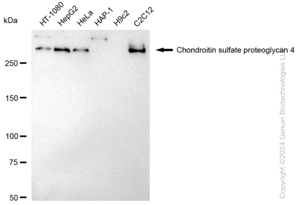
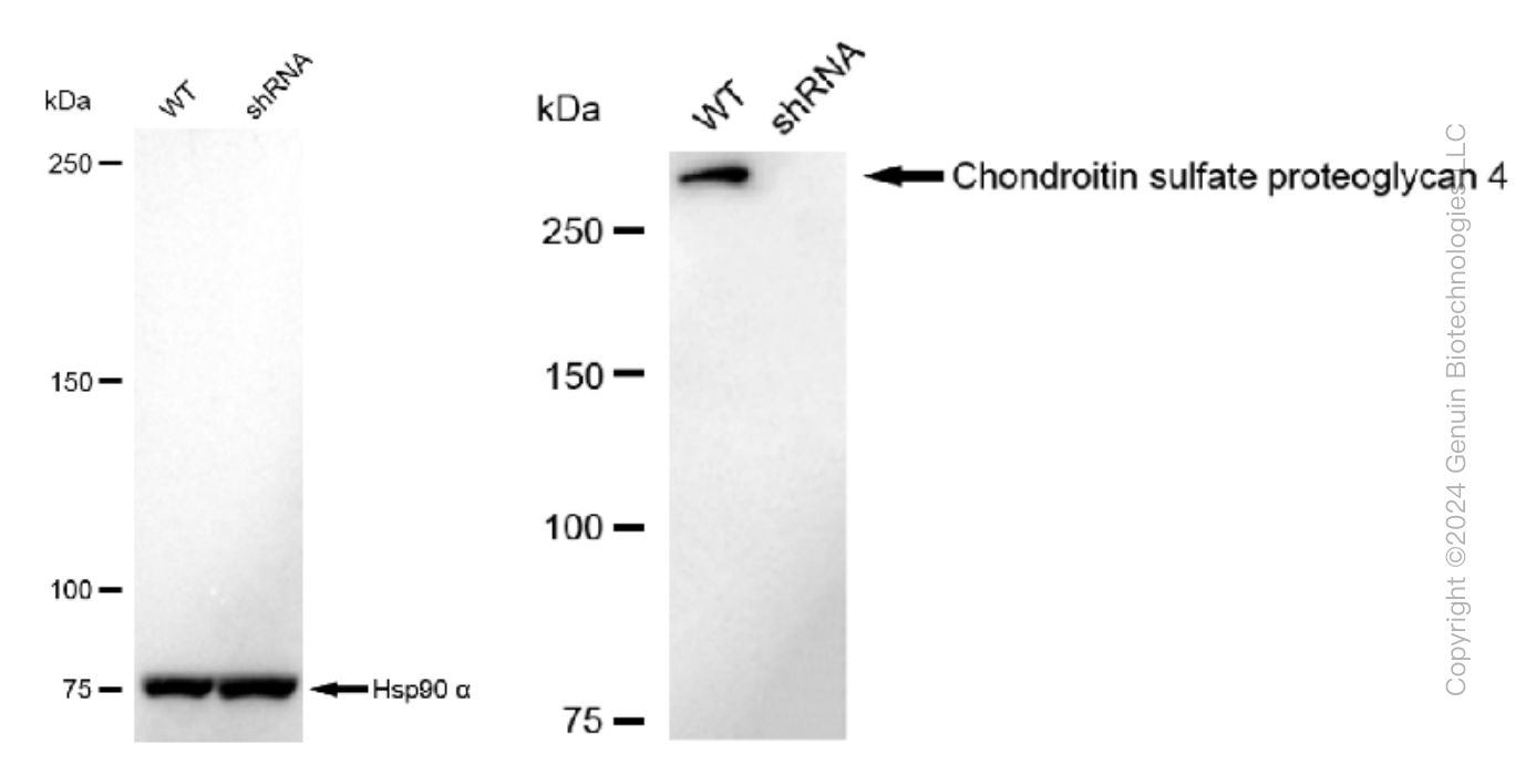
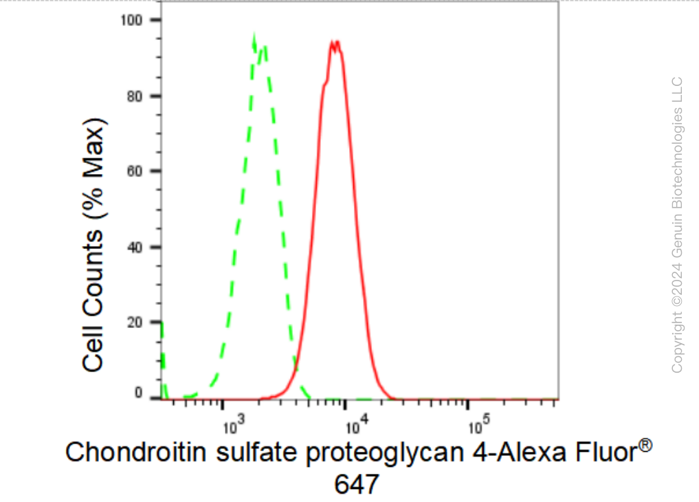
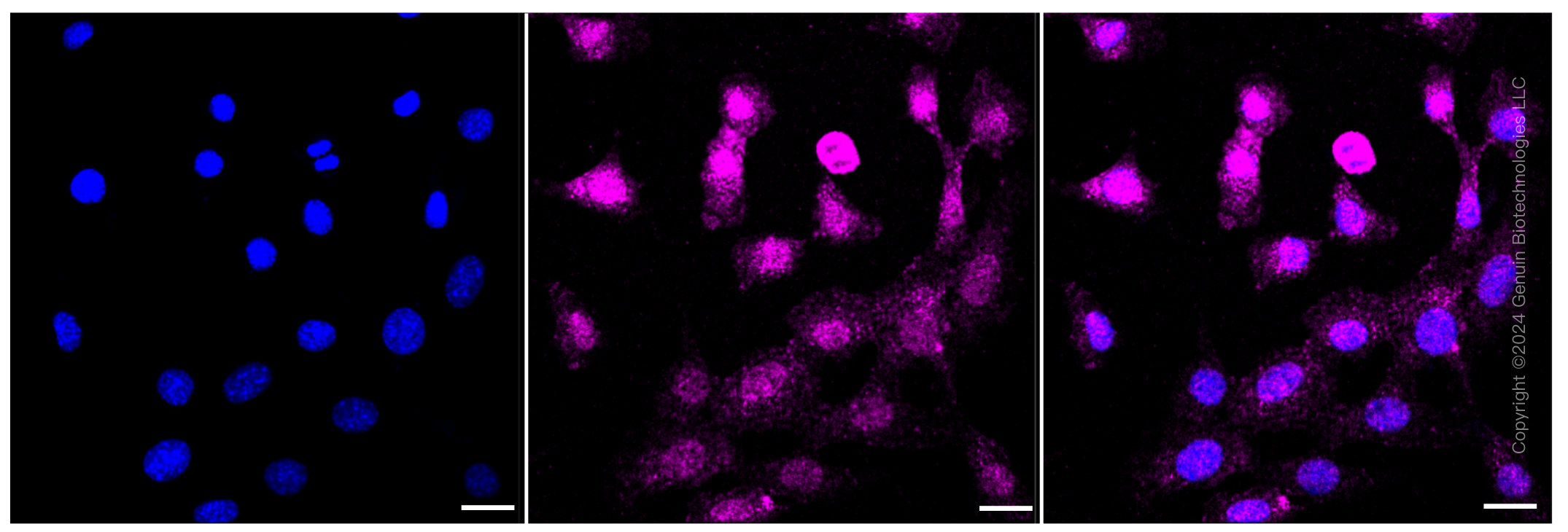
KD-Validated Anti-Chondroitin sulfate proteoglycan 4 Rabbit Monoclonal Antibody
Rabbit monoclonal antibody
- SPECIFICATION
- CITATIONS
- PROTOCOLS
- BACKGROUND

Application
| WB, FC, ICC |
|---|---|
| Primary Accession | Q6UVK1 |
| Reactivity | Human, Mouse |
| Clonality | Monoclonal |
| Isotype | Rabbit IgG |
| Clone Names | 23GB3470 |
| Calculated MW | Predicted, 251 kDa , observed, 280 kDa |
| Gene Name | CSPG4 |
| Aliases | CSPG4; Chondroitin Sulfate Proteoglycan 4; MCSP; Melanoma-Associated Chondroitin Sulfate Proteoglycan; MEL-CSPG; HMW-MAA; CSPG4A ; MCSPG; MSK16; NG2; Chondroitin Sulfate Proteoglycan 4 (Melanoma-Associated); Melanoma Chondroitin Sulfate Proteoglycan; Chondroitin Sulfate Proteoglycan NG2; EC 2.7.8; EC 3.6.3 |
| Immunogen | A synthesized peptide derived from human NG2 |
| Gene ID | 1464 |
|---|---|
| Other Names | Chondroitin sulfate proteoglycan 4, Chondroitin sulfate proteoglycan NG2, Melanoma chondroitin sulfate proteoglycan, Melanoma-associated chondroitin sulfate proteoglycan, CSPG4, MCSP |
| Name | CSPG4 |
|---|---|
| Synonyms | MCSP |
| Function | Proteoglycan playing a role in cell proliferation and migration which stimulates endothelial cells motility during microvascular morphogenesis. May also inhibit neurite outgrowth and growth cone collapse during axon regeneration. Cell surface receptor for collagen alpha 2(VI) which may confer cells ability to migrate on that substrate. Binds through its extracellular N-terminus growth factors, extracellular matrix proteases modulating their activity. May regulate MPP16-dependent degradation and invasion of type I collagen participating in melanoma cells invasion properties. May modulate the plasminogen system by enhancing plasminogen activation and inhibiting angiostatin. Also functions as a signal transducing protein by binding through its cytoplasmic C-terminus scaffolding and signaling proteins. May promote retraction fiber formation and cell polarization through Rho GTPase activation. May stimulate alpha-4, beta-1 integrin-mediated adhesion and spreading by recruiting and activating a signaling cascade through CDC42, ACK1 and BCAR1. May activate FAK and ERK1/ERK2 signaling cascades. |
| Cellular Location | Cell membrane {ECO:0000250|UniProtKB:Q00657}; Single-pass type I membrane protein {ECO:0000250|UniProtKB:Q00657}; Extracellular side {ECO:0000250|UniProtKB:Q00657}. Apical cell membrane {ECO:0000250|UniProtKB:Q00657}; Single-pass type I membrane protein {ECO:0000250|UniProtKB:Q00657}; Extracellular side {ECO:0000250|UniProtKB:Q00657}. Cell projection, lamellipodium membrane {ECO:0000250|UniProtKB:Q00657}; Single-pass type I membrane protein {ECO:0000250|UniProtKB:Q00657}; Extracellular side {ECO:0000250|UniProtKB:Q00657}. Cell surface {ECO:0000250|UniProtKB:Q00657}. Note=Localized at the apical plasma membrane it relocalizes to the lamellipodia of astrocytoma upon phosphorylation by PRKCA. Localizes to the retraction fibers. Localizes to the plasma membrane of oligodendrocytes (By similarity) {ECO:0000250|UniProtKB:Q00657, ECO:0000250|UniProtKB:Q8VHY0} |
| Tissue Location | Detected in fibroblasts (at protein level) (PubMed:36213313). Detected in placenta (at protein level) (PubMed:32337544). Detected in malignant melanoma cells |
Research Areas
Citations (0)

Thousands of laboratories across the world have published research that depended on the performance of antibodies from Abcepta to advance their research. Check out links to articles that cite our products in major peer-reviewed journals, organized by research category.
Submit your citation using an Abcepta antibody to
info@abcepta.com, and receive a free "I Love Antibodies" mug.
info@abcepta.com, and receive a free "I Love Antibodies" mug.

Application Protocols
Provided below are standard protocols that you may find useful for product applications.
Abcepta welcomes feedback from its customers.
If you have used an Abcepta product and would like to share how it has performed, please click on the "Submit Review" button and provide the requested information. Our staff will examine and post your review and contact you if needed.
If you have any additional inquiries please email technical services at tech@abcepta.com.
$ 399.20

$ 149.00
Cat# AGI1333
Ordering Information
United States
AlbaniaAustraliaAustriaBelgiumBosnia & HerzegovinaBrazilBulgariaCanadaCentral AmericaChinaCroatiaCyprusCzech RepublicDenmarkEstoniaFinlandFranceGermanyGreeceHong KongHungaryIcelandIndiaIndonesiaIrelandIsraelItalyJapanLatviaLithuaniaLuxembourgMacedoniaMalaysiaMaltaMexicoNetherlandsNew ZealandNorwayPakistanPolandPortugalRomaniaSerbiaSingaporeSlovakiaSloveniaSouth AfricaSouth KoreaSpainSwedenSwitzerlandTaiwanTurkeyUnited KingdomUnited StatesVietnamWorldwideOthers
USA Headquarters
(888) 735-7227 / (858) 622-0099 or (858) 875-1900
Other Products
Shipping Information
Domestic orders (in stock items)
Shipped out the same day. Orders placed after 1 PM (PST) will ship out the next business day.
International orders
Contact your local distributors
