


 Foundational characteristics of cancer include proliferation, angiogenesis, migration, evasion of apoptosis, and cellular immortality. Find key markers for these cellular processes and antibodies to detect them.
 The SUMOplot™ Analysis Program predicts and scores sumoylation sites in your protein. SUMOylation is a post-translational modification involved in various cellular processes, such as nuclear-cytosolic transport, transcriptional regulation, apoptosis, protein stability, response to stress, and progression through the cell cycle.
 The Autophagy Receptor Motif Plotter predicts and scores autophagy receptor binding sites in your protein. Identifying proteins connected to this pathway is critical to understanding the role of autophagy in physiological as well as pathological processes such as development, differentiation, neurodegenerative diseases, stress, infection, and cancer.
> home > Products > Primary Antibodies > Antibody Collections > KD-Validated Antibodies > KD-Validated Anti-Atg16L1 Rabbit Monoclonal Antibody
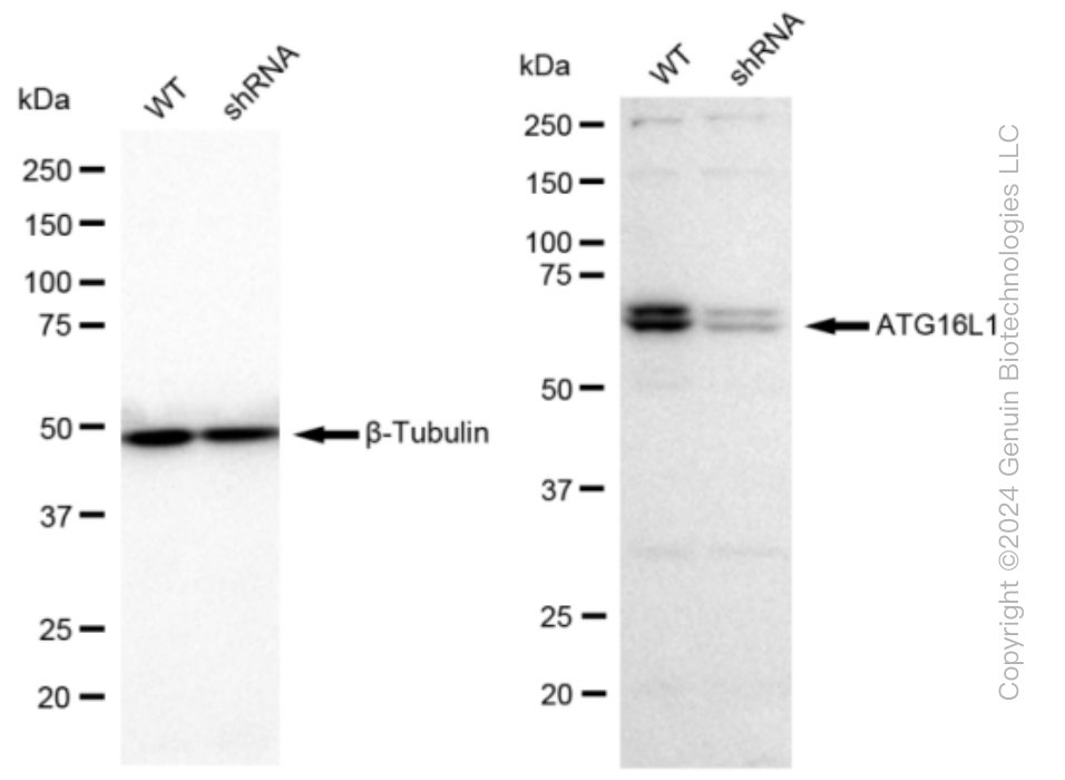
KD-Validated Anti-Atg16L1 Rabbit Monoclonal Antibody
Rabbit monoclonal antibody
- SPECIFICATION
- CITATIONS
- PROTOCOLS
- BACKGROUND

Application
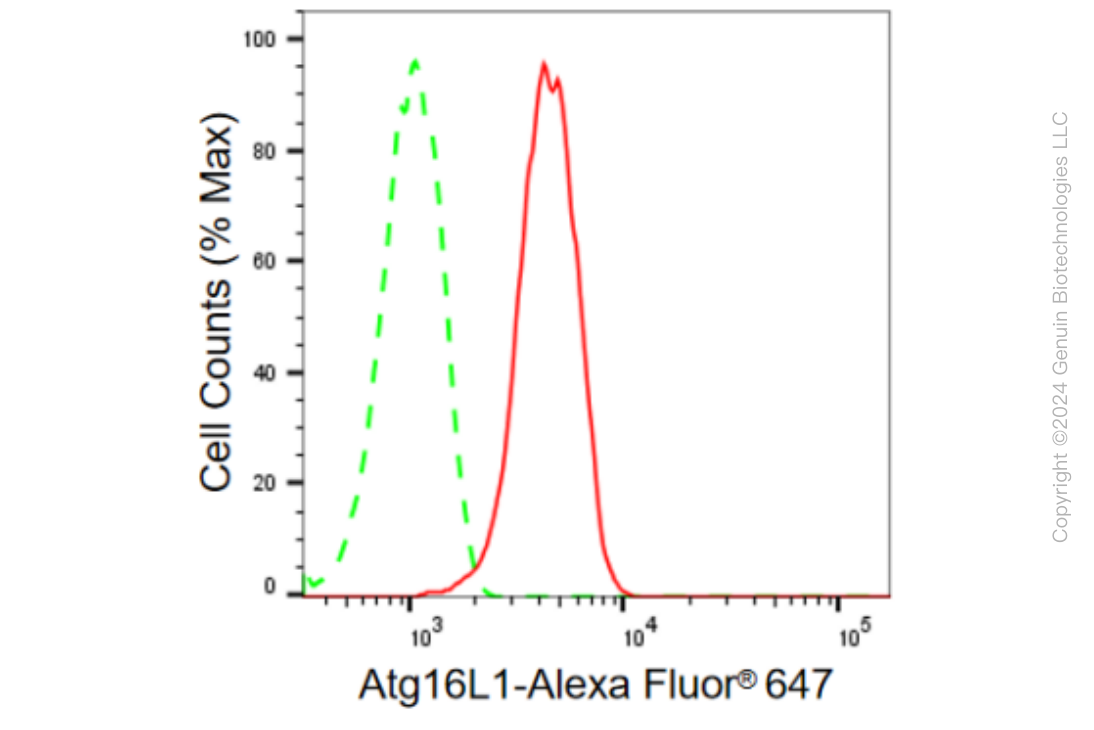
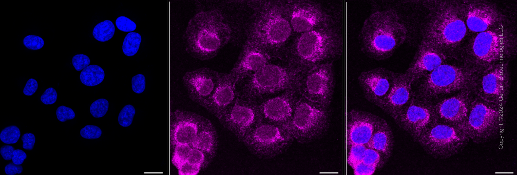
| WB, FC, ICC |
|---|---|
| Primary Accession | Q676U5 |
| Clonality | Monoclonal |
| Isotype | Rabbit IgG |
| Clone Names | 23GB665 Species Reactivity |
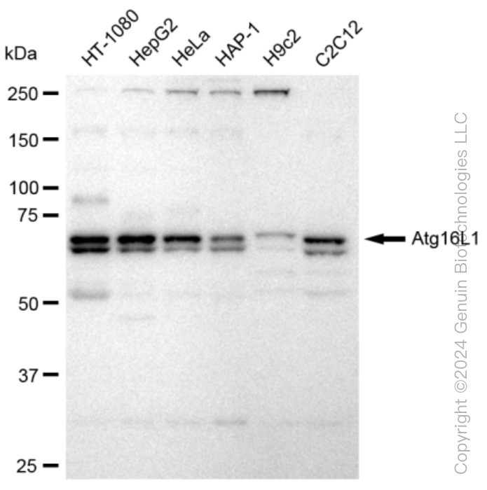
| Calculated MW | Predicted, 68 kDa , observed, 68 kDa |
| Gene Name | ATG16L1 |
| Aliases | ATG16L1; Autophagy Related 16 Like 1; ATG16A; APG16L; WDR30; Autophagy-Related Protein 16-1; FLJ10035; ATG16L; ATG16 Autophagy Related 16-Like 1 (S. Cerevisiae); ATG16 Autophagy Related 16-Like (S. Cerevisiae); APG16 Autophagy 16-Like (S. Cerevisiae); ATG16 Autophagy Related 16-Like 1; WD Repeat Domain 30; APG16-Like 1; APG16L Beta; IBD104C, Cysteine Peptidase; ATG4 Autophagy Related 4 Homolog C; AUT-Like 1, Cysteine; Endopeptidase; APG4 Autophagy 4 Homolog C; EC 3.4.22.-; EC 3.4.22; APG4-C |
| Immunogen | A synthesized peptide derived from human ATG16L1 |
| Gene ID | 55054 |
|---|---|
| Other Names | Autophagy-related protein 16-1, APG16-like 1, ATG16L1 {ECO:0000303|PubMed:17200669, ECO:0000312|HGNC:HGNC:21498} |
| Name | ATG16L1 {ECO:0000303|PubMed:17200669, ECO:0000312|HGNC:HGNC:21498} |
|---|---|
| Function | Plays an essential role in both canonical and non-canonical autophagy: interacts with ATG12-ATG5 to mediate the lipidation to ATG8 family proteins (MAP1LC3A, MAP1LC3B, MAP1LC3C, GABARAPL1, GABARAPL2 and GABARAP) (PubMed:23376921, PubMed:23392225, PubMed:24553140, PubMed:24954904, PubMed:27273576, PubMed:29317426, PubMed:30778222, PubMed:33909989). Acts as a molecular hub, coordinating autophagy pathways via distinct domains that support either canonical or non- canonical signaling (PubMed:29317426, PubMed:30778222). During canonical autophagy, interacts with ATG12-ATG5 to mediate the conjugation of phosphatidylethanolamine (PE) to ATG8 proteins, to produce a membrane-bound activated form of ATG8 (PubMed:23376921, PubMed:23392225, PubMed:24553140, PubMed:24954904, PubMed:27273576). Thereby, controls the elongation of the nascent autophagosomal membrane (PubMed:23376921, PubMed:23392225, PubMed:24553140, PubMed:24954904, PubMed:27273576). As part of the ATG8 conjugation system with ATG5 and ATG12, required for recruitment of LRRK2 to stressed lysosomes and induction of LRRK2 kinase activity in response to lysosomal stress (By similarity). Also involved in non-canonical autophagy, a parallel pathway involving conjugation of ATG8 proteins to single membranes at endolysosomal compartments, probably by catalyzing conjugation of phosphatidylserine (PS) to ATG8 (PubMed:33909989). Non-canonical autophagy plays a key role in epithelial cells to limit lethal infection by influenza A (IAV) virus (By similarity). Regulates mitochondrial antiviral signaling (MAVS)-dependent type I interferon (IFN-I) production (PubMed:22749352, PubMed:25645662). Negatively regulates NOD1- and NOD2-driven inflammatory cytokine response (PubMed:24238340). Instead, promotes an autophagy-dependent antibacterial pathway together with NOD1 or NOD2 (PubMed:20637199). Plays a role in regulating morphology and function of Paneth cell (PubMed:18849966). |
| Cellular Location | Cytoplasm. Preautophagosomal structure membrane; Peripheral membrane protein. Endosome membrane; Peripheral membrane protein. Lysosome membrane; Peripheral membrane protein. Note=Recruited to omegasomes membranes by WIPI2 (By similarity). Omegasomes are endoplasmic reticulum connected strutures at the origin of preautophagosomal structures (By similarity). Localized to preautophagosomal structure (PAS) where it is involved in the membrane targeting of ATG5 (By similarity). Also localizes to discrete punctae along the ciliary axoneme (By similarity). Upon activation of non-canonical autophagy, recruited to single-membrane endolysosomal compartments (PubMed:29317426). Under starved conditions, the ATG12-ATG5-ATG16L1 complex is translocated to phagophores driven by RAB33B (PubMed:32960676). {ECO:0000250|UniProtKB:Q8C0J2, ECO:0000269|PubMed:29317426, ECO:0000269|PubMed:32960676} |
Research Areas
Citations (0)

Thousands of laboratories across the world have published research that depended on the performance of antibodies from Abcepta to advance their research. Check out links to articles that cite our products in major peer-reviewed journals, organized by research category.
Submit your citation using an Abcepta antibody to
info@abcepta.com, and receive a free "I Love Antibodies" mug.
info@abcepta.com, and receive a free "I Love Antibodies" mug.

Application Protocols
Provided below are standard protocols that you may find useful for product applications.
Abcepta welcomes feedback from its customers.
If you have used an Abcepta product and would like to share how it has performed, please click on the "Submit Review" button and provide the requested information. Our staff will examine and post your review and contact you if needed.
If you have any additional inquiries please email technical services at tech@abcepta.com.
$ 399.20

$ 149.00
Cat# AGI1364
Ordering Information
United States
AlbaniaAustraliaAustriaBelgiumBosnia & HerzegovinaBrazilBulgariaCanadaCentral AmericaChinaCroatiaCyprusCzech RepublicDenmarkEstoniaFinlandFranceGermanyGreeceHong KongHungaryIcelandIndiaIndonesiaIrelandIsraelItalyJapanLatviaLithuaniaLuxembourgMacedoniaMalaysiaMaltaMexicoNetherlandsNew ZealandNorwayPakistanPolandPortugalRomaniaSerbiaSingaporeSlovakiaSloveniaSouth AfricaSouth KoreaSpainSwedenSwitzerlandTaiwanTurkeyUnited KingdomUnited StatesVietnamWorldwideOthers
USA Headquarters
(888) 735-7227 / (858) 622-0099 or (858) 875-1900
Other Products
Shipping Information
Domestic orders (in stock items)
Shipped out the same day. Orders placed after 1 PM (PST) will ship out the next business day.
International orders
Contact your local distributors
