


 Foundational characteristics of cancer include proliferation, angiogenesis, migration, evasion of apoptosis, and cellular immortality. Find key markers for these cellular processes and antibodies to detect them.
 The SUMOplot™ Analysis Program predicts and scores sumoylation sites in your protein. SUMOylation is a post-translational modification involved in various cellular processes, such as nuclear-cytosolic transport, transcriptional regulation, apoptosis, protein stability, response to stress, and progression through the cell cycle.
 The Autophagy Receptor Motif Plotter predicts and scores autophagy receptor binding sites in your protein. Identifying proteins connected to this pathway is critical to understanding the role of autophagy in physiological as well as pathological processes such as development, differentiation, neurodegenerative diseases, stress, infection, and cancer.
> home > Products > Primary Antibodies > Antibody Collections > Head neck negative > KD-Validated Anti-SBDS Ribosome MatuRation Factor Rabbit Monoclonal Antibody
KD-Validated Anti-SBDS Ribosome MatuRation Factor Rabbit Monoclonal Antibody
Rabbit monoclonal antibody
- SPECIFICATION
- CITATIONS
- PROTOCOLS
- BACKGROUND

Application
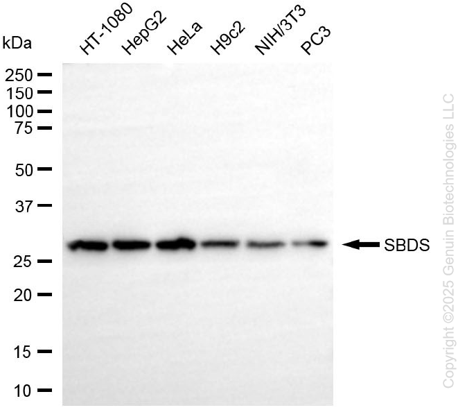
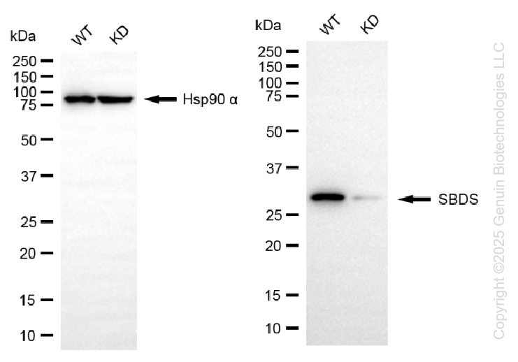
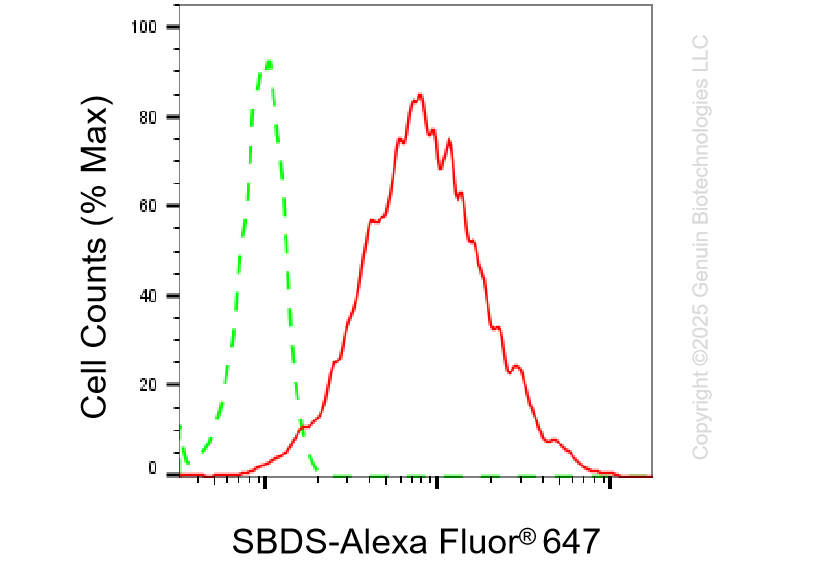
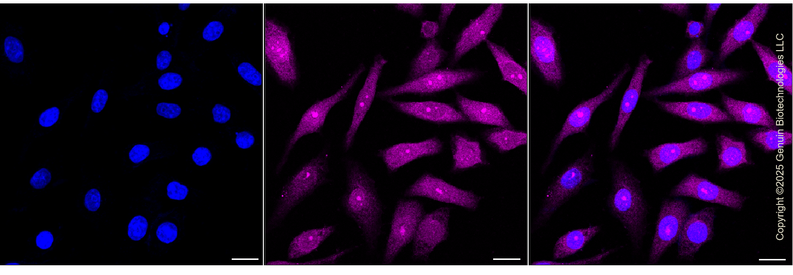
| WB, FC, ICC |
|---|---|
| Primary Accession | Q9Y3A5 |
| Reactivity | Rat, Human, Mouse |
| Clonality | Monoclonal |
| Isotype | Rabbit IgG |
| Clone Names | 25GB3180 |
| Calculated MW | Predicted, 29 kDa; observed, 29 kDa |
| Gene Name | SBDS |
| Aliases | SBDS; SBDS Ribosome Maturation Factor; CGI-97; SWDS; SDO1; SDS; SBDS, Ribosome Assembly Guanine Nucleotide Exchange Factor; Ribosome Maturation Protein SBDS; FLJ10917; Shwachman-Bodian-Diamond Syndrome Protein; Shwachman-Bodian-Diamond Syndrome |
| Immunogen | A synthesized peptide derived from human SBDS |
| Gene ID | 51119 |
|---|---|
| Other Names | Ribosome maturation protein SBDS, Shwachman-Bodian-Diamond syndrome protein, SBDS |
| Name | SBDS |
|---|---|
| Function | Required for the assembly of mature ribosomes and ribosome biogenesis. Together with EFL1, triggers the GTP-dependent release of EIF6 from 60S pre-ribosomes in the cytoplasm, thereby activating ribosomes for translation competence by allowing 80S ribosome assembly and facilitating EIF6 recycling to the nucleus, where it is required for 60S rRNA processing and nuclear export. Required for normal levels of protein synthesis. May play a role in cellular stress resistance. May play a role in cellular response to DNA damage. May play a role in cell proliferation. |
| Cellular Location | Cytoplasm. Nucleus, nucleolus. Nucleus, nucleoplasm. Cytoplasm, cytoskeleton, spindle. Note=Primarily detected in the cytoplasm, and at low levels in nucleus and nucleolus (PubMed:17475909, PubMed:19602484). Detected in the nucleolus during G1 and G2 phase of the cell cycle, and diffusely distributed in the nucleus during S phase. Detected at the mitotic spindle. Colocalizes with the microtubule organizing center during interphase (PubMed:19759903). |
| Tissue Location | Widely expressed. |
Research Areas
Citations (0)

Thousands of laboratories across the world have published research that depended on the performance of antibodies from Abcepta to advance their research. Check out links to articles that cite our products in major peer-reviewed journals, organized by research category.
Submit your citation using an Abcepta antibody to
info@abcepta.com, and receive a free "I Love Antibodies" mug.
info@abcepta.com, and receive a free "I Love Antibodies" mug.

Application Protocols
Provided below are standard protocols that you may find useful for product applications.
Abcepta welcomes feedback from its customers.
If you have used an Abcepta product and would like to share how it has performed, please click on the "Submit Review" button and provide the requested information. Our staff will examine and post your review and contact you if needed.
If you have any additional inquiries please email technical services at tech@abcepta.com.
$ 399.20

$ 149.00
Cat# AGI1373
Ordering Information
United States
AlbaniaAustraliaAustriaBelgiumBosnia & HerzegovinaBrazilBulgariaCanadaCentral AmericaChinaCroatiaCyprusCzech RepublicDenmarkEstoniaFinlandFranceGermanyGreeceHong KongHungaryIcelandIndiaIndonesiaIrelandIsraelItalyJapanLatviaLithuaniaLuxembourgMacedoniaMalaysiaMaltaMexicoNetherlandsNew ZealandNorwayPakistanPolandPortugalRomaniaSerbiaSingaporeSlovakiaSloveniaSouth AfricaSouth KoreaSpainSwedenSwitzerlandTaiwanTurkeyUnited KingdomUnited StatesVietnamWorldwideOthers
USA Headquarters
(888) 735-7227 / (858) 622-0099 or (858) 875-1900
Other Products
Shipping Information
Domestic orders (in stock items)
Shipped out the same day. Orders placed after 1 PM (PST) will ship out the next business day.
International orders
Contact your local distributors
