


 Foundational characteristics of cancer include proliferation, angiogenesis, migration, evasion of apoptosis, and cellular immortality. Find key markers for these cellular processes and antibodies to detect them.
 The SUMOplot™ Analysis Program predicts and scores sumoylation sites in your protein. SUMOylation is a post-translational modification involved in various cellular processes, such as nuclear-cytosolic transport, transcriptional regulation, apoptosis, protein stability, response to stress, and progression through the cell cycle.
 The Autophagy Receptor Motif Plotter predicts and scores autophagy receptor binding sites in your protein. Identifying proteins connected to this pathway is critical to understanding the role of autophagy in physiological as well as pathological processes such as development, differentiation, neurodegenerative diseases, stress, infection, and cancer.
> home > Products > Primary Antibodies > Antibody Collections > KD-Validated Antibodies > KD-Validated Anti-AKT3 Mouse Monoclonal Antibody
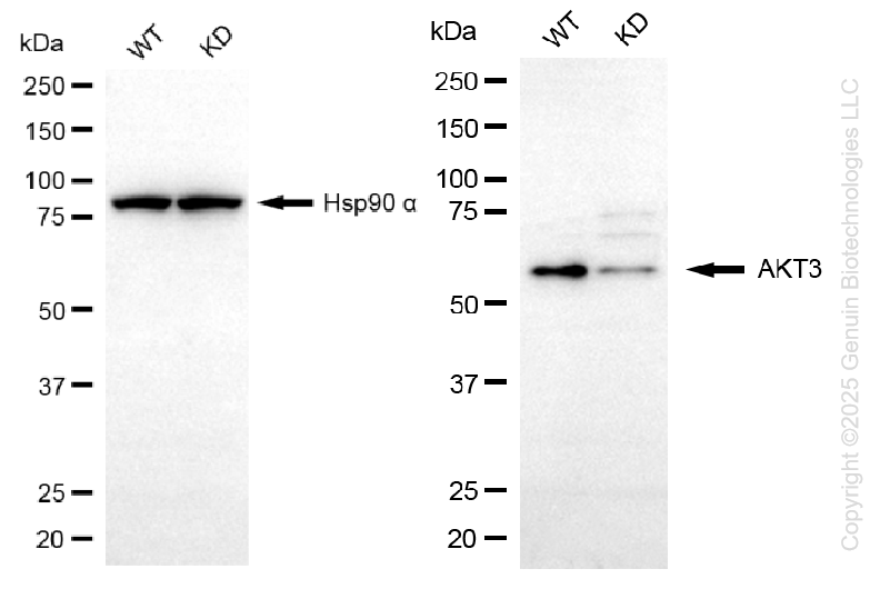
KD-Validated Anti-AKT3 Mouse Monoclonal Antibody
Mouse monoclonal antibody
- SPECIFICATION
- CITATIONS
- PROTOCOLS
- BACKGROUND

Application
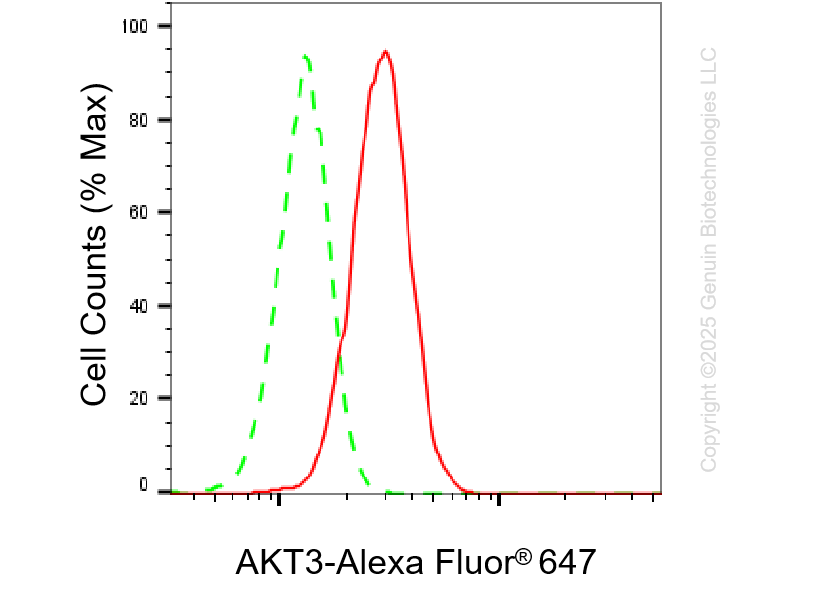
| WB, FC |
|---|---|
| Primary Accession | Q9Y243 |
| Reactivity | Rat, Human, Mouse |
| Clonality | Monoclonal |
| Isotype | Mouse IgG3 |
| Clone Names | 25GB3680 |
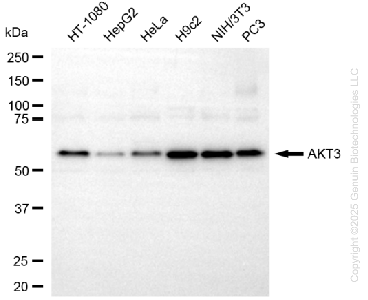
| Calculated MW | Predicted, 56 kDa; observed, 56 kDa |
| Gene Name | AKT3 |
| Aliases | AKT3; AKT Serine/Threonine Kinase 3; PKBG; RAC-Gamma; PRKBG; RAC-Gamma Serine/Threonine-Protein Kinase; RAC-PK-Gamma; EC 2.7.11.1; PKB Gamma; STK-2; V-Akt Murine Thymoma Viral Oncogene Homolog 3 (Protein Kinase B, Gamma); V-Akt Murine Thymoma Viral Oncogene Homolog 3; RAC-Gamma Serine/Threonine Protein Kinase; Protein Kinase B, Gamma; Protein Kinase B Gamma; Protein Kinase Akt-3; PKB-GAMMA; EC 2.7.11; MPPH2; MPPH |
| Immunogen | Recombinant protein of human AKT3 |
| Gene ID | 10000 |
|---|---|
| Other Names | RAC-gamma serine/threonine-protein kinase, 2.7.11.1, Protein kinase Akt-3, Protein kinase B gamma, PKB gamma, RAC-PK-gamma, STK-2, AKT3, PKBG |
| Name | AKT3 |
|---|---|
| Synonyms | PKBG |
| Function | AKT3 is one of 3 closely related serine/threonine-protein kinases (AKT1, AKT2 and AKT3) called the AKT kinase, and which regulate many processes including metabolism, proliferation, cell survival, growth and angiogenesis. This is mediated through serine and/or threonine phosphorylation of a range of downstream substrates. Over 100 substrate candidates have been reported so far, but for most of them, no isoform specificity has been reported. AKT3 is the least studied AKT isoform. It plays an important role in brain development and is crucial for the viability of malignant glioma cells. AKT3 isoform may also be the key molecule in up-regulation and down-regulation of MMP13 via IL13. Required for the coordination of mitochondrial biogenesis with growth factor-induced increases in cellular energy demands. Down- regulation by RNA interference reduces the expression of the phosphorylated form of BAD, resulting in the induction of caspase- dependent apoptosis. |
| Cellular Location | Nucleus. Cytoplasm. Membrane; Peripheral membrane protein Note=Membrane-associated after cell stimulation leading to its translocation |
| Tissue Location | In adult tissues, it is highly expressed in brain, lung and kidney, but weakly in heart, testis and liver. In fetal tissues, it is highly expressed in heart, liver and brain and not at all in kidney |
Research Areas
Citations (0)

Thousands of laboratories across the world have published research that depended on the performance of antibodies from Abcepta to advance their research. Check out links to articles that cite our products in major peer-reviewed journals, organized by research category.
Submit your citation using an Abcepta antibody to
info@abcepta.com, and receive a free "I Love Antibodies" mug.
info@abcepta.com, and receive a free "I Love Antibodies" mug.

Application Protocols
Provided below are standard protocols that you may find useful for product applications.
Abcepta welcomes feedback from its customers.
If you have used an Abcepta product and would like to share how it has performed, please click on the "Submit Review" button and provide the requested information. Our staff will examine and post your review and contact you if needed.
If you have any additional inquiries please email technical services at tech@abcepta.com.
$ 399.20

$ 74.50

⚠️ Limit: 5 vials per order for discount.
Cat# AGI1441
Ordering Information
United States
AlbaniaAustraliaAustriaBelgiumBosnia & HerzegovinaBrazilBulgariaCanadaCentral AmericaChinaCroatiaCyprusCzech RepublicDenmarkEstoniaFinlandFranceGermanyGreeceHong KongHungaryIcelandIndiaIndonesiaIrelandIsraelItalyJapanLatviaLithuaniaLuxembourgMacedoniaMalaysiaMaltaMexicoNetherlandsNew ZealandNorwayPakistanPolandPortugalRomaniaSerbiaSingaporeSlovakiaSloveniaSouth AfricaSouth KoreaSpainSwedenSwitzerlandTaiwanTurkeyUnited KingdomUnited StatesVietnamWorldwideOthers
USA Headquarters
(888) 735-7227 / (858) 622-0099 or (858) 875-1900
Other Products
Shipping Information
Domestic orders (in stock items)
Shipped out the same day. Orders placed after 1 PM (PST) will ship out the next business day.
International orders
Contact your local distributors
