


 Foundational characteristics of cancer include proliferation, angiogenesis, migration, evasion of apoptosis, and cellular immortality. Find key markers for these cellular processes and antibodies to detect them.
 The SUMOplot™ Analysis Program predicts and scores sumoylation sites in your protein. SUMOylation is a post-translational modification involved in various cellular processes, such as nuclear-cytosolic transport, transcriptional regulation, apoptosis, protein stability, response to stress, and progression through the cell cycle.
 The Autophagy Receptor Motif Plotter predicts and scores autophagy receptor binding sites in your protein. Identifying proteins connected to this pathway is critical to understanding the role of autophagy in physiological as well as pathological processes such as development, differentiation, neurodegenerative diseases, stress, infection, and cancer.
> home > Products > Primary Antibodies > Antibody Collections > KD-Validated Antibodies > KD-Validated Anti-GOPC Rabbit Monoclonal Antibody
KD-Validated Anti-GOPC Rabbit Monoclonal Antibody
Rabbit monoclonal antibody
- SPECIFICATION
- CITATIONS
- PROTOCOLS
- BACKGROUND

Application
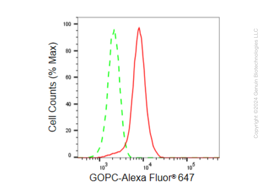
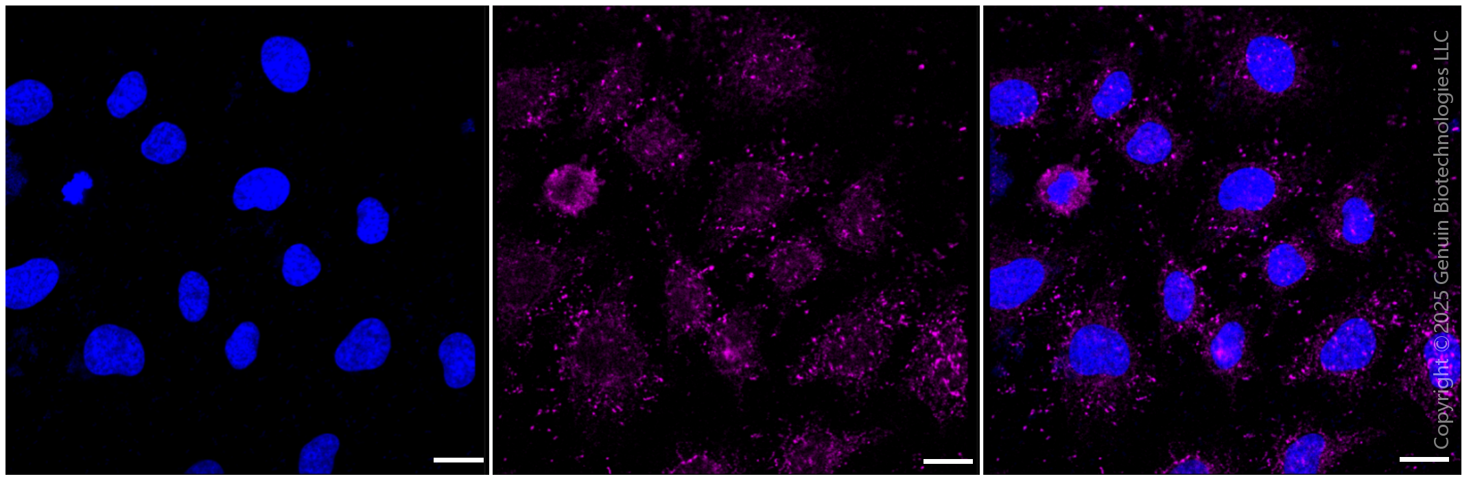
| WB, FC, ICC |
|---|---|
| Primary Accession | Q9HD26 |
| Reactivity | Rat, Human, Mouse |
| Clonality | Monoclonal |
| Isotype | Rabbit IgG |
| Clone Names | 23GB5870 |
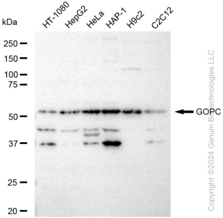
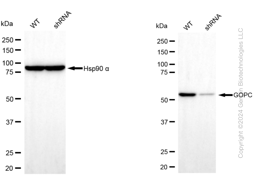
| Calculated MW | Predicted, 51 kDa , observed , 51 kDa |
| Gene Name | GOPC |
| Aliases | GOPC; Golgi Associated PDZ And Coiled-Coil Motif Containing; PIST; FIG; CAL; DJ94G16.2; GOPC1; Golgi-Associated PDZ And Coiled-Coil Motif-Containing Protein; PDZ Protein Interacting Specifically With TC10; CFTR-Associated Ligand; Fused In Glioblastoma; PDZ/Coiled-Coil Domain Binding Partner For The Rho-Family GTPase TC10; Golgi-Associated PDZ And Coiled-Coil Motif Containing Protein; DJ94G16.2 PIST |
| Immunogen | A synthesized peptide derived from human PIST |
| Gene ID | 57120 |
|---|---|
| Other Names | Golgi-associated PDZ and coiled-coil motif-containing protein {ECO:0000312|HGNC:HGNC:17643}, CFTR-associated ligand, Fused in glioblastoma, PDZ protein interacting specifically with TC10, PIST, GOPC (HGNC:17643) |
| Name | GOPC (HGNC:17643) |
|---|---|
| Function | Plays a role in intracellular protein trafficking and degradation (PubMed:11707463, PubMed:14570915, PubMed:15358775). May regulate CFTR chloride currents and acid-induced ASIC3 currents by modulating cell surface expression of both channels (By similarity). May also regulate the intracellular trafficking of the ADR1B receptor (PubMed:15358775). May play a role in autophagy (By similarity). Together with MARCHF2 mediates the ubiquitination and lysosomal degradation of CFTR (PubMed:23818989). Overexpression results in CFTR intracellular retention and lysosomaldegradation in the lysosomes (PubMed:11707463, PubMed:14570915). |
| Cellular Location | Cytoplasm. Golgi apparatus membrane; Peripheral membrane protein. Golgi apparatus, trans-Golgi network membrane; Peripheral membrane protein Synapse. Postsynaptic density. Cell projection, dendrite. Note=Enriched in synaptosomal and postsynaptic densities (PSD) fractions. Expressed in cell bodies and dendrites of Purkinje cells. Localized at the trans-Golgi network (TGN) of spermatids and the medulla of round spermatides. |
| Tissue Location | Ubiquitously expressed. |
Research Areas
Citations (0)

Thousands of laboratories across the world have published research that depended on the performance of antibodies from Abcepta to advance their research. Check out links to articles that cite our products in major peer-reviewed journals, organized by research category.
Submit your citation using an Abcepta antibody to
info@abcepta.com, and receive a free "I Love Antibodies" mug.
info@abcepta.com, and receive a free "I Love Antibodies" mug.

Application Protocols
Provided below are standard protocols that you may find useful for product applications.
Abcepta welcomes feedback from its customers.
If you have used an Abcepta product and would like to share how it has performed, please click on the "Submit Review" button and provide the requested information. Our staff will examine and post your review and contact you if needed.
If you have any additional inquiries please email technical services at tech@abcepta.com.
$ 399.20

$ 149.00
Cat# AGI1544
Ordering Information
United States
AlbaniaAustraliaAustriaBelgiumBosnia & HerzegovinaBrazilBulgariaCanadaCentral AmericaChinaCroatiaCyprusCzech RepublicDenmarkEstoniaFinlandFranceGermanyGreeceHong KongHungaryIcelandIndiaIndonesiaIrelandIsraelItalyJapanLatviaLithuaniaLuxembourgMacedoniaMalaysiaMaltaMexicoNetherlandsNew ZealandNorwayPakistanPolandPortugalRomaniaSerbiaSingaporeSlovakiaSloveniaSouth AfricaSouth KoreaSpainSwedenSwitzerlandTaiwanTurkeyUnited KingdomUnited StatesVietnamWorldwideOthers
USA Headquarters
(888) 735-7227 / (858) 622-0099 or (858) 875-1900
Other Products
Shipping Information
Domestic orders (in stock items)
Shipped out the same day. Orders placed after 1 PM (PST) will ship out the next business day.
International orders
Contact your local distributors
