


 Foundational characteristics of cancer include proliferation, angiogenesis, migration, evasion of apoptosis, and cellular immortality. Find key markers for these cellular processes and antibodies to detect them.
 The SUMOplot™ Analysis Program predicts and scores sumoylation sites in your protein. SUMOylation is a post-translational modification involved in various cellular processes, such as nuclear-cytosolic transport, transcriptional regulation, apoptosis, protein stability, response to stress, and progression through the cell cycle.
 The Autophagy Receptor Motif Plotter predicts and scores autophagy receptor binding sites in your protein. Identifying proteins connected to this pathway is critical to understanding the role of autophagy in physiological as well as pathological processes such as development, differentiation, neurodegenerative diseases, stress, infection, and cancer.
> home > Products > Primary Antibodies > Antibody Collections > KD-Validated Antibodies > KD-Validated Anti-KSR1 Rabbit Monoclonal Antibody
KD-Validated Anti-KSR1 Rabbit Monoclonal Antibody
Rabbit monoclonal antibody

- SPECIFICATION
- CITATIONS
- PROTOCOLS
- BACKGROUND

Application
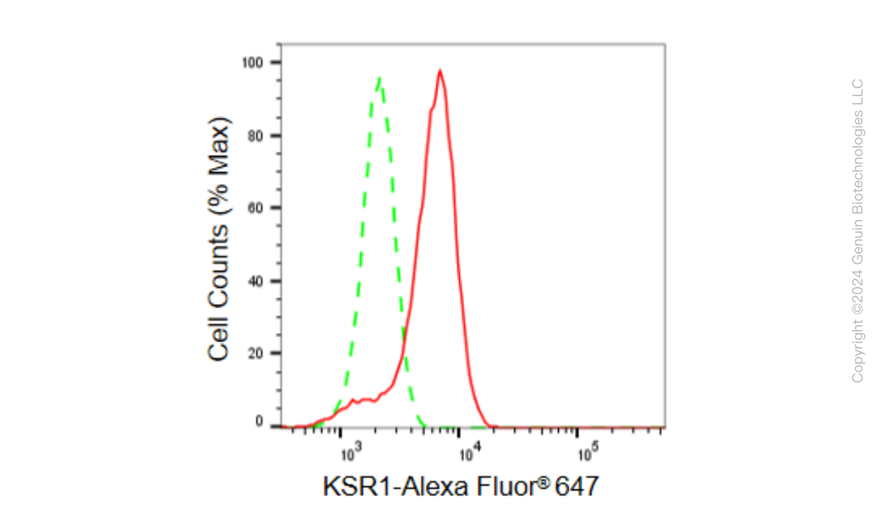
| WB, FC |
|---|---|
| Primary Accession | Q8IVT5 |
| Reactivity | Rat, Human |
| Clonality | Monoclonal |
| Isotype | Rabbit IgG |
| Clone Names | 23GB6290 |
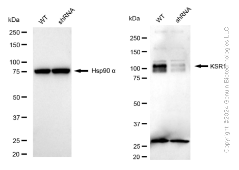
| Calculated MW | Predicted, 102 kDa , observed , 120 kDa |
| Gene Name | KSR1 |
| Aliases | KSR1; Kinase Suppressor Of Ras 1; RSU2; KSR; Kinase Suppressor Of Ras; EC 2.7.11.1 |
| Immunogen | A synthesized peptide derived from human KSR1 |
| Gene ID | 8844 |
|---|---|
| Other Names | Kinase suppressor of Ras 1, 2.7.11.1, KSR1, KSR |
| Name | KSR1 |
|---|---|
| Synonyms | KSR |
| Function | Part of a multiprotein signaling complex which promotes phosphorylation of Raf family members and activation of downstream MAP kinases (By similarity). Independently of its kinase activity, acts as MAP2K1/MEK1 and MAP2K2/MEK2-dependent allosteric activator of BRAF; upon binding to MAP2K1/MEK1 or MAP2K2/MEK2, dimerizes with BRAF and promotes BRAF-mediated phosphorylation of MAP2K1/MEK1 and/or MAP2K2/MEK2 (PubMed:29433126). Promotes activation of MAPK1 and/or MAPK3, both in response to EGF and to cAMP (By similarity). Its kinase activity is unsure (By similarity). Some protein kinase activity has been detected in vitro, however the physiological relevance of this activity is unknown (By similarity). |
| Cellular Location | Cytoplasm. Membrane; Peripheral membrane protein. Cell membrane {ECO:0000250|UniProtKB:Q61097}; Peripheral membrane protein {ECO:0000250|UniProtKB:Q61097}. Cell projection, ruffle membrane {ECO:0000250|UniProtKB:Q61097}. Endoplasmic reticulum membrane. Note=In unstimulated cells, where the phosphorylated form is bound to a 14-3-3 protein, sequestration in the cytoplasm occurs. Following growth factor treatment, the protein is free for membrane translocation, and it moves from the cytoplasm to the cell periphery. |
Research Areas
Citations (0)

Thousands of laboratories across the world have published research that depended on the performance of antibodies from Abcepta to advance their research. Check out links to articles that cite our products in major peer-reviewed journals, organized by research category.
Submit your citation using an Abcepta antibody to
info@abcepta.com, and receive a free "I Love Antibodies" mug.
info@abcepta.com, and receive a free "I Love Antibodies" mug.

Application Protocols
Provided below are standard protocols that you may find useful for product applications.
Abcepta welcomes feedback from its customers.
If you have used an Abcepta product and would like to share how it has performed, please click on the "Submit Review" button and provide the requested information. Our staff will examine and post your review and contact you if needed.
If you have any additional inquiries please email technical services at tech@abcepta.com.
$ 399.20

$ 149.00
Cat# AGI1561
Ordering Information
United States
AlbaniaAustraliaAustriaBelgiumBosnia & HerzegovinaBrazilBulgariaCanadaCentral AmericaChinaCroatiaCyprusCzech RepublicDenmarkEstoniaFinlandFranceGermanyGreeceHong KongHungaryIcelandIndiaIndonesiaIrelandIsraelItalyJapanLatviaLithuaniaLuxembourgMacedoniaMalaysiaMaltaMexicoNetherlandsNew ZealandNorwayPakistanPolandPortugalRomaniaSerbiaSingaporeSlovakiaSloveniaSouth AfricaSouth KoreaSpainSwedenSwitzerlandTaiwanTurkeyUnited KingdomUnited StatesVietnamWorldwideOthers
USA Headquarters
(888) 735-7227 / (858) 622-0099 or (858) 875-1900
Other Products
Shipping Information
Domestic orders (in stock items)
Shipped out the same day. Orders placed after 1 PM (PST) will ship out the next business day.
International orders
Contact your local distributors
