


 Foundational characteristics of cancer include proliferation, angiogenesis, migration, evasion of apoptosis, and cellular immortality. Find key markers for these cellular processes and antibodies to detect them.
 The SUMOplot™ Analysis Program predicts and scores sumoylation sites in your protein. SUMOylation is a post-translational modification involved in various cellular processes, such as nuclear-cytosolic transport, transcriptional regulation, apoptosis, protein stability, response to stress, and progression through the cell cycle.
 The Autophagy Receptor Motif Plotter predicts and scores autophagy receptor binding sites in your protein. Identifying proteins connected to this pathway is critical to understanding the role of autophagy in physiological as well as pathological processes such as development, differentiation, neurodegenerative diseases, stress, infection, and cancer.
> home > Products > Primary Antibodies > Antibody Collections > KD-Validated Antibodies > KD-Validated Anti-KHSRP Rabbit Monoclonal Antibody
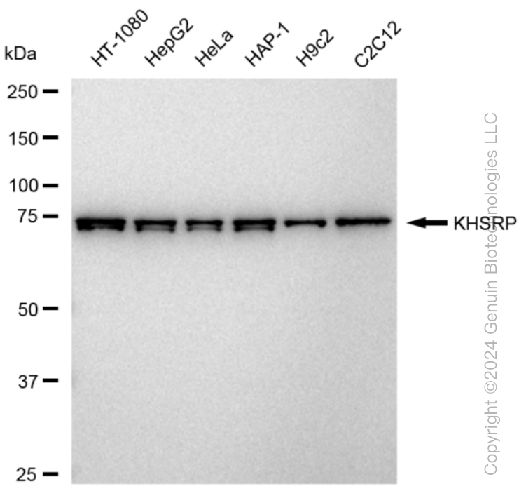
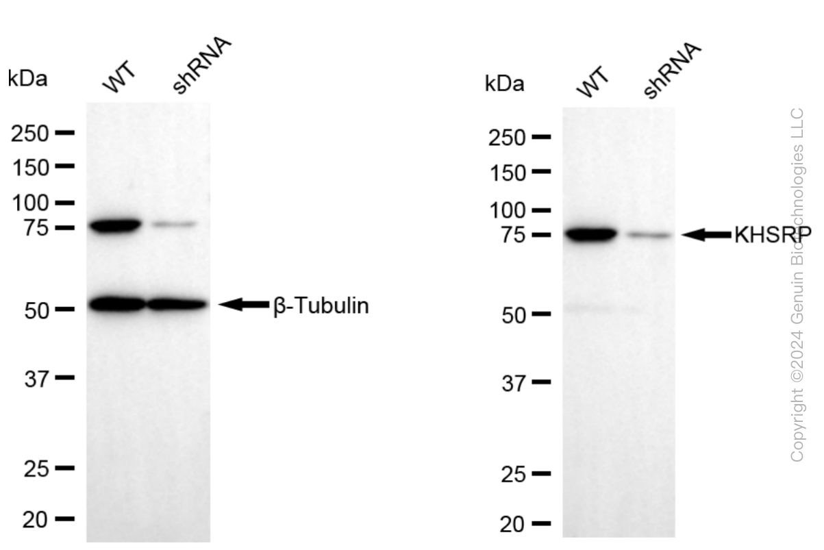
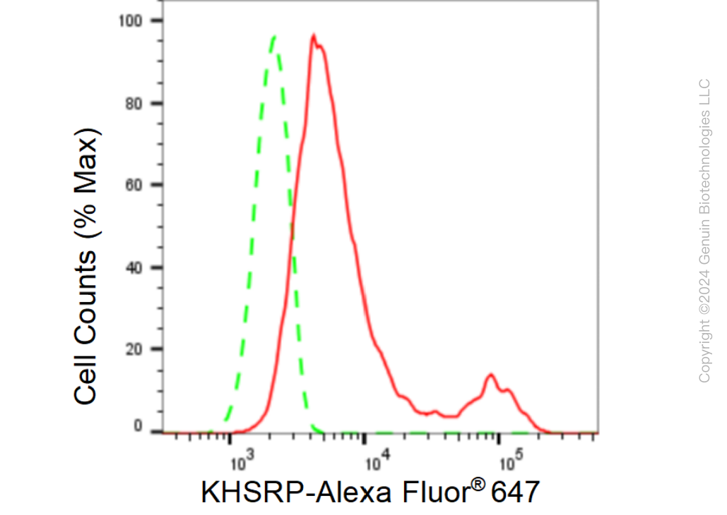
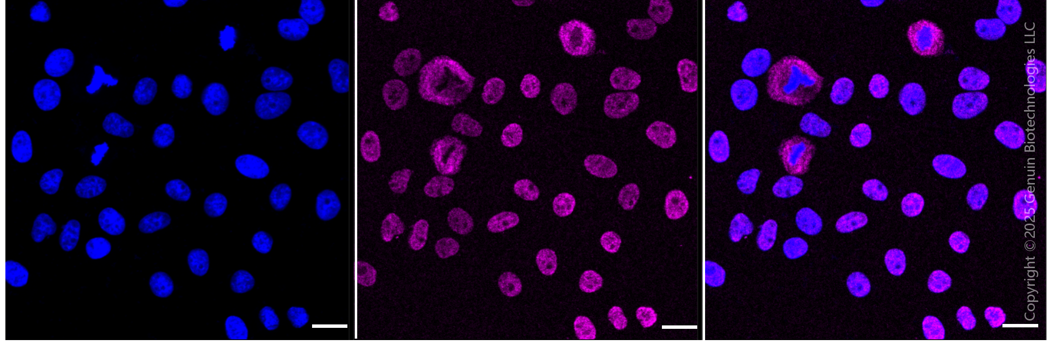
KD-Validated Anti-KHSRP Rabbit Monoclonal Antibody
Rabbit monoclonal antibody
- SPECIFICATION
- CITATIONS
- PROTOCOLS
- BACKGROUND

Application
| WB, FC, ICC |
|---|---|
| Primary Accession | Q92945 |
| Reactivity | Rat, Human, Mouse |
| Clonality | Monoclonal |
| Isotype | Rabbit IgG |
| Clone Names | 23GB6435 |
| Calculated MW | Predicted, 73 kDa , observed , 73 kDa |
| Gene Name | KHSRP |
| Aliases | KHSRP; KH-Type Splicing Regulatory Protein; FUBP2; KSRP; FBP2; Far Upstream Element-Binding Protein; FUSE-Binding Protein; P75; Epididymis Secretory Sperm Binding Protein; KH Type-Splicing Regulatory Protein; FUSE Binding Protein 2 |
| Immunogen | A synthesized peptide derived from human KHSRP |
| Gene ID | 8570 |
|---|---|
| Other Names | Far upstream element-binding protein 2, FUSE-binding protein 2, KH type-splicing regulatory protein, KSRP, p75, KHSRP, FUBP2 |
| Name | KHSRP |
|---|---|
| Synonyms | FUBP2 |
| Function | Binds to the dendritic targeting element and may play a role in mRNA trafficking (By similarity). Part of a ternary complex that binds to the downstream control sequence (DCS) of the pre-mRNA. Mediates exon inclusion in transcripts that are subject to tissue- specific alternative splicing. May interact with single-stranded DNA from the far-upstream element (FUSE). May activate gene expression. Also involved in degradation of inherently unstable mRNAs that contain AU-rich elements (AREs) in their 3'-UTR, possibly by recruiting degradation machinery to ARE-containing mRNAs. |
| Cellular Location | Nucleus. Cytoplasm. Note=A small proportion is also found in the cytoplasm of neuronal cell bodies and dendrites. |
| Tissue Location | Detected in neural and non-neural cell lines. |
Research Areas
Citations (0)

Thousands of laboratories across the world have published research that depended on the performance of antibodies from Abcepta to advance their research. Check out links to articles that cite our products in major peer-reviewed journals, organized by research category.
Submit your citation using an Abcepta antibody to
info@abcepta.com, and receive a free "I Love Antibodies" mug.
info@abcepta.com, and receive a free "I Love Antibodies" mug.

Application Protocols
Provided below are standard protocols that you may find useful for product applications.
Abcepta welcomes feedback from its customers.
If you have used an Abcepta product and would like to share how it has performed, please click on the "Submit Review" button and provide the requested information. Our staff will examine and post your review and contact you if needed.
If you have any additional inquiries please email technical services at tech@abcepta.com.
$ 399.20

$ 149.00
Cat# AGI1566
Ordering Information
United States
AlbaniaAustraliaAustriaBelgiumBosnia & HerzegovinaBrazilBulgariaCanadaCentral AmericaChinaCroatiaCyprusCzech RepublicDenmarkEstoniaFinlandFranceGermanyGreeceHong KongHungaryIcelandIndiaIndonesiaIrelandIsraelItalyJapanLatviaLithuaniaLuxembourgMacedoniaMalaysiaMaltaMexicoNetherlandsNew ZealandNorwayPakistanPolandPortugalRomaniaSerbiaSingaporeSlovakiaSloveniaSouth AfricaSouth KoreaSpainSwedenSwitzerlandTaiwanTurkeyUnited KingdomUnited StatesVietnamWorldwideOthers
USA Headquarters
(888) 735-7227 / (858) 622-0099 or (858) 875-1900
Other Products
Shipping Information
Domestic orders (in stock items)
Shipped out the same day. Orders placed after 1 PM (PST) will ship out the next business day.
International orders
Contact your local distributors
