


 Foundational characteristics of cancer include proliferation, angiogenesis, migration, evasion of apoptosis, and cellular immortality. Find key markers for these cellular processes and antibodies to detect them.
 The SUMOplot™ Analysis Program predicts and scores sumoylation sites in your protein. SUMOylation is a post-translational modification involved in various cellular processes, such as nuclear-cytosolic transport, transcriptional regulation, apoptosis, protein stability, response to stress, and progression through the cell cycle.
 The Autophagy Receptor Motif Plotter predicts and scores autophagy receptor binding sites in your protein. Identifying proteins connected to this pathway is critical to understanding the role of autophagy in physiological as well as pathological processes such as development, differentiation, neurodegenerative diseases, stress, infection, and cancer.
> home > Products > Primary Antibodies > Antibody Collections > KD-Validated Antibodies > KD-Validated Anti-IL13 receptor alpha 1 Rabbit Monoclonal Antibody
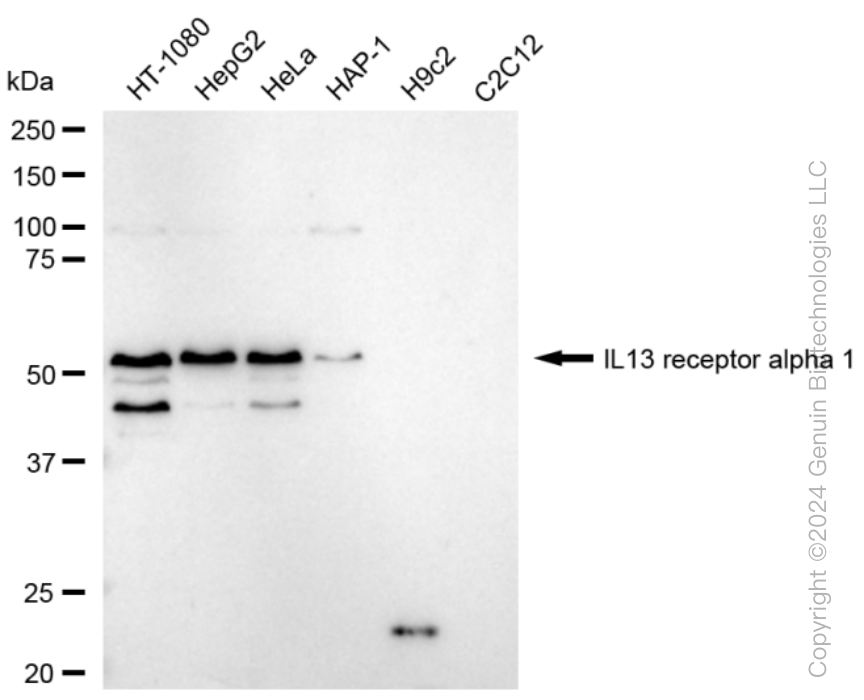
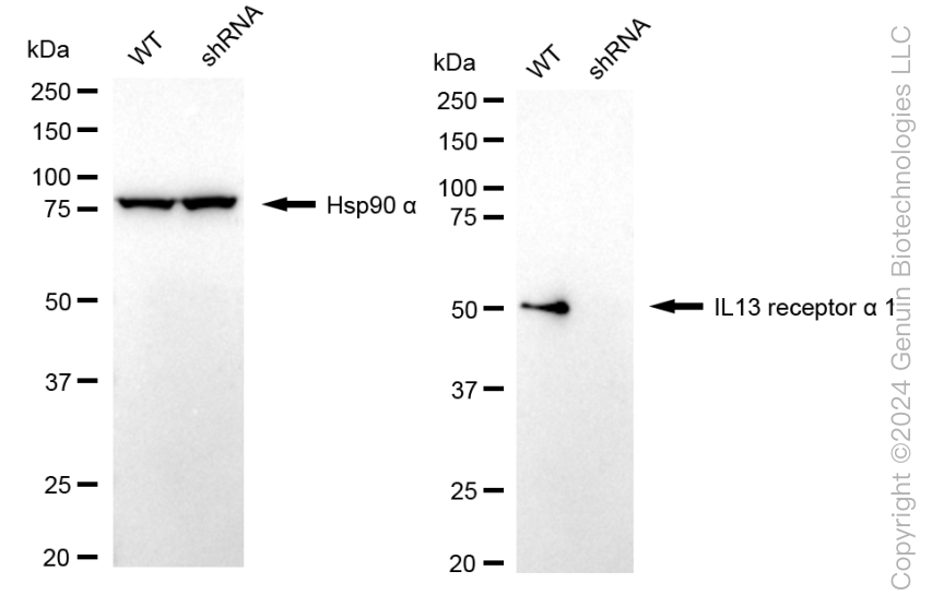
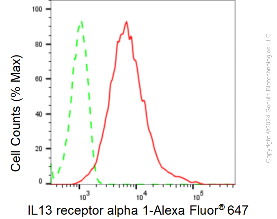
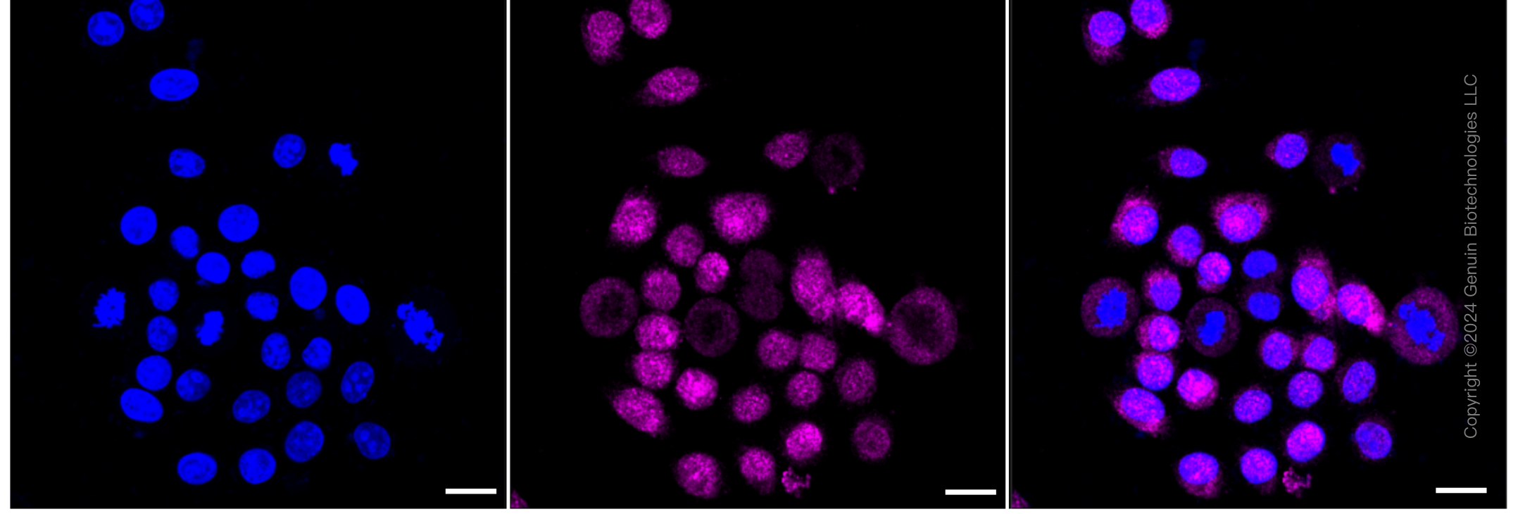
KD-Validated Anti-IL13 receptor alpha 1 Rabbit Monoclonal Antibody
Rabbit monoclonal antibody
- SPECIFICATION
- CITATIONS
- PROTOCOLS
- BACKGROUND

Application
| WB, FC, ICC |
|---|---|
| Primary Accession | P78552 |
| Reactivity | Human |
| Clonality | Monoclonal |
| Isotype | Rabbit IgG |
| Clone Names | 23GB6655 |
| Calculated MW | Predicted, 49 kDa , observed , 50 kDa |
| Gene Name | IL13RA1 |
| Aliases | IL13RA1; Interleukin 13 Receptor Subunit Alpha 1; CD213a1 Antigen; IL-13Ra; CD213a1; NR4; Interleukin-13 Receptor Subunit Alpha-1; Interleukin 13 Receptor, Alpha 1; IL-13 Receptor Subunit Alpha-1; IL13 Receptor Alpha-1 Chain; Cancer/Testis Antigen 19; IL-13R Subunit Alpha-1; CT19; IL-13R-Alpha-1; IL-13RA1; IL13RA; IL13R |
| Immunogen | A synthesized peptide derived from human IL13 receptor alpha 1 |
| Gene ID | 3597 |
|---|---|
| Other Names | Interleukin-13 receptor subunit alpha-1, IL-13 receptor subunit alpha-1, IL-13R subunit alpha-1, IL-13R-alpha-1, IL-13RA1, Cancer/testis antigen 19, CT19, CD213a1, IL13RA1, IL13R, IL13RA |
| Name | IL13RA1 |
|---|---|
| Synonyms | IL13R, IL13RA |
| Function | Binds with low affinity to interleukin-13 (IL13). Together with IL4RA can form a functional receptor for IL13. Also serves as an alternate accessory protein to the common cytokine receptor gamma chain for interleukin-4 (IL4) signaling, but cannot replace the function of IL2RG in allowing enhanced interleukin-2 (IL2) binding activity. |
| Cellular Location | Membrane; Single-pass type I membrane protein. |
| Tissue Location | Ubiquitous. Highest levels in heart, liver, skeletal muscle and ovary; lowest levels in brain, lung and kidney Also found in B-cells, T-cells and endothelial cells |
Research Areas
Citations (0)

Thousands of laboratories across the world have published research that depended on the performance of antibodies from Abcepta to advance their research. Check out links to articles that cite our products in major peer-reviewed journals, organized by research category.
Submit your citation using an Abcepta antibody to
info@abcepta.com, and receive a free "I Love Antibodies" mug.
info@abcepta.com, and receive a free "I Love Antibodies" mug.

Application Protocols
Provided below are standard protocols that you may find useful for product applications.
Abcepta welcomes feedback from its customers.
If you have used an Abcepta product and would like to share how it has performed, please click on the "Submit Review" button and provide the requested information. Our staff will examine and post your review and contact you if needed.
If you have any additional inquiries please email technical services at tech@abcepta.com.
$ 399.20

$ 149.00
Cat# AGI1576
Ordering Information
United States
AlbaniaAustraliaAustriaBelgiumBosnia & HerzegovinaBrazilBulgariaCanadaCentral AmericaChinaCroatiaCyprusCzech RepublicDenmarkEstoniaFinlandFranceGermanyGreeceHong KongHungaryIcelandIndiaIndonesiaIrelandIsraelItalyJapanLatviaLithuaniaLuxembourgMacedoniaMalaysiaMaltaMexicoNetherlandsNew ZealandNorwayPakistanPolandPortugalRomaniaSerbiaSingaporeSlovakiaSloveniaSouth AfricaSouth KoreaSpainSwedenSwitzerlandTaiwanTurkeyUnited KingdomUnited StatesVietnamWorldwideOthers
USA Headquarters
(888) 735-7227 / (858) 622-0099 or (858) 875-1900
Other Products
Shipping Information
Domestic orders (in stock items)
Shipped out the same day. Orders placed after 1 PM (PST) will ship out the next business day.
International orders
Contact your local distributors
