


 Foundational characteristics of cancer include proliferation, angiogenesis, migration, evasion of apoptosis, and cellular immortality. Find key markers for these cellular processes and antibodies to detect them.
 The SUMOplot™ Analysis Program predicts and scores sumoylation sites in your protein. SUMOylation is a post-translational modification involved in various cellular processes, such as nuclear-cytosolic transport, transcriptional regulation, apoptosis, protein stability, response to stress, and progression through the cell cycle.
 The Autophagy Receptor Motif Plotter predicts and scores autophagy receptor binding sites in your protein. Identifying proteins connected to this pathway is critical to understanding the role of autophagy in physiological as well as pathological processes such as development, differentiation, neurodegenerative diseases, stress, infection, and cancer.
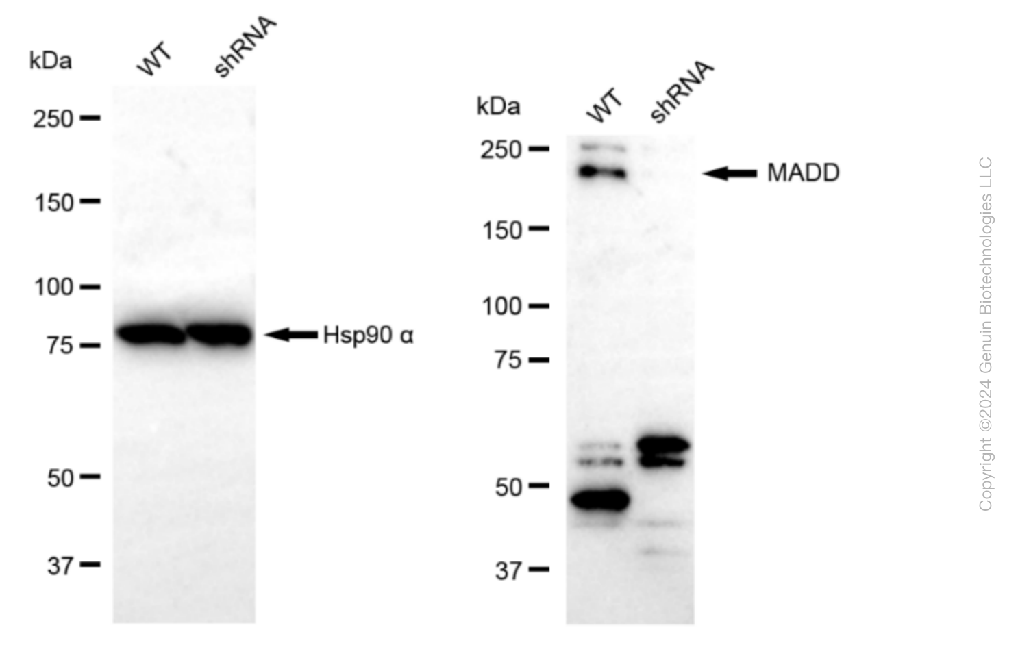
> home > Products > Primary Antibodies > Antibody Collections > KD-Validated Antibodies > KD-Validated Anti-MAP Kinase Activating Death Domain Rabbit Monoclonal Antibody
KD-Validated Anti-MAP Kinase Activating Death Domain Rabbit Monoclonal Antibody
Rabbit monoclonal antibody
- SPECIFICATION
- CITATIONS
- PROTOCOLS
- BACKGROUND

Application
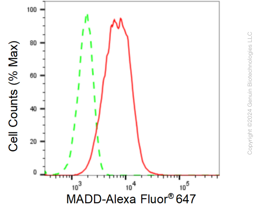
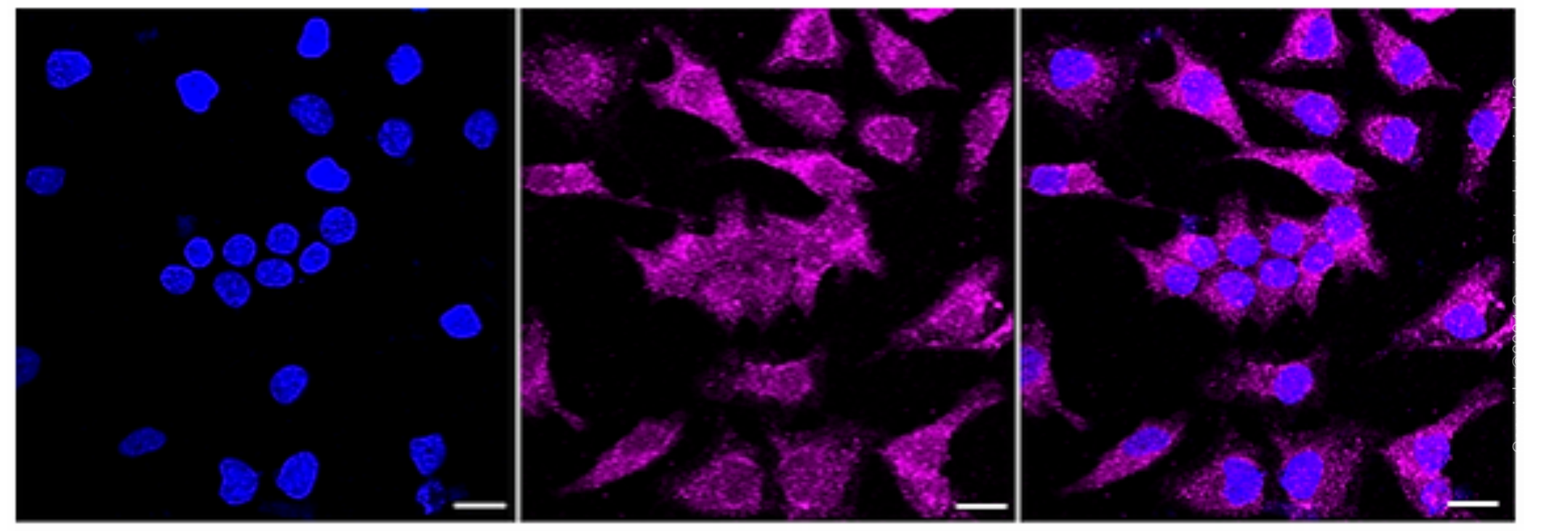
| WB, FC, ICC |
|---|---|
| Primary Accession | Q8WXG6 |
| Reactivity | Rat, Human, Mouse |
| Clonality | Monoclonal |
| Isotype | Rabbit IgG |
| Clone Names | 24GB110 |
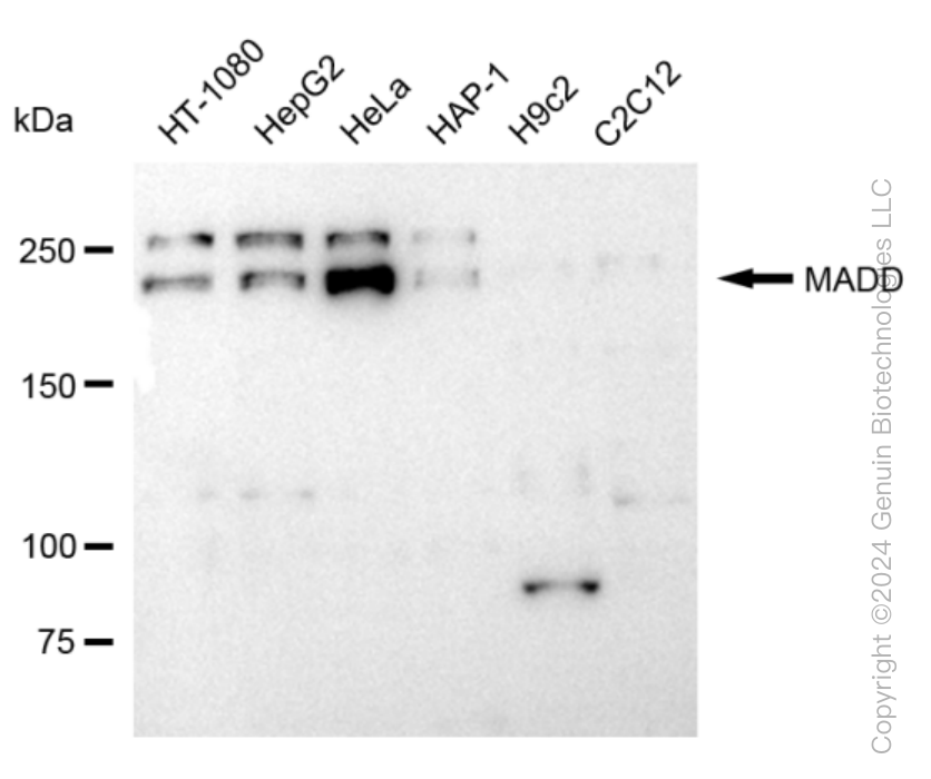
| Calculated MW | Predicted, 183 kDa, observed , 230 kDa |
| Gene Name | MADD |
| Aliases | MAP Kinase Activating Death Domain; DENN; Differentially Expressed In Normal And Neoplastic Cells; KIAA0358; RAB3GEP; IG20; MAP Kinase-Activating Death Domain Protein; Insuloma-Glucagonoma Protein 20; Insulinoma Glucagonoma Clone 20; Rab3 GDP/GTP Exchange Protein; Rab3 GDP/GTP Exchange Factor; RabGEF; MAP-Kinase Activating Death Domain; NEDDISH; Rab3GEP; DEEAH |
| Immunogen | A synthesized peptide derived from human DENN |
| Gene ID | 8567 |
|---|---|
| Other Names | MAP kinase-activating death domain protein, Differentially expressed in normal and neoplastic cells, Insulinoma glucagonoma clone 20, Rab3 GDP/GTP exchange factor, RabGEF, Rab3 GDP/GTP exchange protein, Rab3GEP, MADD {ECO:0000312|EMBL:AAB57735.1, ECO:0000312|HGNC:HGNC:6766} |
| Name | MADD {ECO:0000312|EMBL:AAB57735.1, ECO:0000312|HGNC:HGNC:6766} |
|---|---|
| Function | Guanyl-nucleotide exchange factor that regulates small GTPases of the Rab family (PubMed:18559336, PubMed:20937701). Converts GDP-bound inactive form of RAB27A and RAB27B to the GTP-bound active forms (PubMed:18559336, PubMed:20937701). Converts GDP-bound inactive form of RAB3A, RAB3C and RAB3D to the GTP-bound active forms, GTPases involved in synaptic vesicle exocytosis and vesicle secretion (By similarity). Plays a role in synaptic vesicle formation and in vesicle trafficking at the neuromuscular junction (By similarity). Involved in up-regulating a post-docking step of synaptic exocytosis in central synapses (By similarity). Probably by binding to the motor proteins KIF1B and KIF1A, mediates motor-dependent transport of GTP-RAB3A- positive vesicles to the presynaptic nerve terminals (By similarity). Plays a role in TNFA-mediated activation of the MAPK pathway, including ERK1/2 (PubMed:32761064). May link TNFRSF1A with MAP kinase activation (PubMed:9115275). May be involved in the regulation of TNFA-induced apoptosis (PubMed:11577081, PubMed:32761064). |
| Cellular Location | Cell membrane. Cytoplasm. Cell projection, axon {ECO:0000250|UniProtKB:Q80U28} |
| Tissue Location | Expressed in testis, ovary, brain and heart (PubMed:8988362). Expressed in spleen, thymus, prostate, testis, ovary, small instestine and colon (PubMed:9115275). Expressed in liver (PubMed:9796103). [Isoform 2]: Expressed in the brain, breast, kidney, lung, ovary, pancreas, testis, uterus, stomach and thyroid [Isoform 4]: Expressed in the brain, breast, kidney, lung, ovary, pancreas, testis, uterus, stomach and thyroid [Isoform 6]: Not detected in the brain, breast, kidney, lung, ovary, pancreas, testis, uterus, stomach and thyroid |
Research Areas
Citations (0)

Thousands of laboratories across the world have published research that depended on the performance of antibodies from Abcepta to advance their research. Check out links to articles that cite our products in major peer-reviewed journals, organized by research category.
Submit your citation using an Abcepta antibody to
info@abcepta.com, and receive a free "I Love Antibodies" mug.
info@abcepta.com, and receive a free "I Love Antibodies" mug.

Application Protocols
Provided below are standard protocols that you may find useful for product applications.
Abcepta welcomes feedback from its customers.
If you have used an Abcepta product and would like to share how it has performed, please click on the "Submit Review" button and provide the requested information. Our staff will examine and post your review and contact you if needed.
If you have any additional inquiries please email technical services at tech@abcepta.com.
$ 399.20

$ 149.00
Cat# AGI1587
Ordering Information
United States
AlbaniaAustraliaAustriaBelgiumBosnia & HerzegovinaBrazilBulgariaCanadaCentral AmericaChinaCroatiaCyprusCzech RepublicDenmarkEstoniaFinlandFranceGermanyGreeceHong KongHungaryIcelandIndiaIndonesiaIrelandIsraelItalyJapanLatviaLithuaniaLuxembourgMacedoniaMalaysiaMaltaMexicoNetherlandsNew ZealandNorwayPakistanPolandPortugalRomaniaSerbiaSingaporeSlovakiaSloveniaSouth AfricaSouth KoreaSpainSwedenSwitzerlandTaiwanTurkeyUnited KingdomUnited StatesVietnamWorldwideOthers
USA Headquarters
(888) 735-7227 / (858) 622-0099 or (858) 875-1900
Other Products
Shipping Information
Domestic orders (in stock items)
Shipped out the same day. Orders placed after 1 PM (PST) will ship out the next business day.
International orders
Contact your local distributors
