


 Foundational characteristics of cancer include proliferation, angiogenesis, migration, evasion of apoptosis, and cellular immortality. Find key markers for these cellular processes and antibodies to detect them.
 The SUMOplot™ Analysis Program predicts and scores sumoylation sites in your protein. SUMOylation is a post-translational modification involved in various cellular processes, such as nuclear-cytosolic transport, transcriptional regulation, apoptosis, protein stability, response to stress, and progression through the cell cycle.
 The Autophagy Receptor Motif Plotter predicts and scores autophagy receptor binding sites in your protein. Identifying proteins connected to this pathway is critical to understanding the role of autophagy in physiological as well as pathological processes such as development, differentiation, neurodegenerative diseases, stress, infection, and cancer.
> home > Products > Primary Antibodies > Antibody Collections > SARS-Human interaction partners > KD-Validated Anti-Ribophorin I Rabbit Monoclonal Antibody
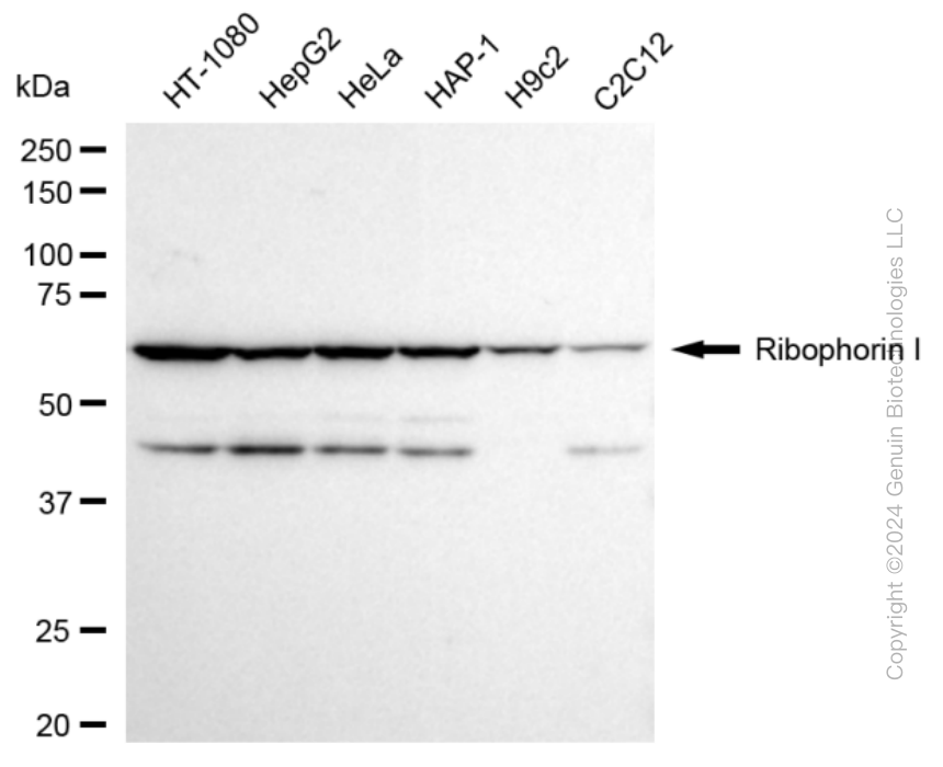
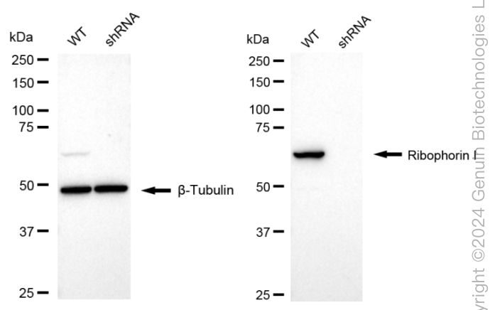
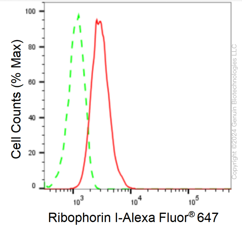
KD-Validated Anti-Ribophorin I Rabbit Monoclonal Antibody
Rabbit monoclonal antibody
- SPECIFICATION
- CITATIONS
- PROTOCOLS
- BACKGROUND

Application
| WB, FC |
|---|---|
| Primary Accession | P04843 |
| Reactivity | Rat, Human, Mouse |
| Clonality | Monoclonal |
| Isotype | Rabbit IgG |
| Clone Names | 24GB1090 |
| Calculated MW | Predicted, 69 kDa , observed , 69 kDa |
| Gene Name | RPN1 |
| Aliases | Ribophorin I; OST1; Dolichyl-Diphosphooligosaccharide--Protein Glycosyltransferase Subunit 1; Oligosaccharyltransferase Complex Subunit (Non-Catalytic); Ribophorin-1; RPN-I; Dolichyl-Diphosphooligosaccharide--Protein Glycosyltransferase 67 KDa Subunit; Dolichyl-Diphosphooligosaccharide-Protein Glycosyltransferase 67 KDa Subunit; Oligosaccharyltransferase 1 Homolog (S. Cerevisiae); Oligosaccharyltransferase 1 Homolog; EC 2.4.1.119; RBPH1 |
| Immunogen | A synthesized peptide derived from human PRibophorin I |
| Gene ID | 6184 |
|---|---|
| Other Names | Dolichyl-diphosphooligosaccharide--protein glycosyltransferase subunit 1, Dolichyl-diphosphooligosaccharide--protein glycosyltransferase 67 kDa subunit, Ribophorin I, RPN-I, Ribophorin-1, RPN1 (HGNC:10381) |
| Name | RPN1 (HGNC:10381) |
|---|---|
| Function | Subunit of the oligosaccharyl transferase (OST) complex that catalyzes the initial transfer of a defined glycan (Glc(3)Man(9)GlcNAc(2) in eukaryotes) from the lipid carrier dolichol- pyrophosphate to an asparagine residue within an Asn-X-Ser/Thr consensus motif in nascent polypeptide chains, the first step in protein N-glycosylation (PubMed:31831667). N-glycosylation occurs cotranslationally and the complex associates with the Sec61 complex at the channel-forming translocon complex that mediates protein translocation across the endoplasmic reticulum (ER). All subunits are required for a maximal enzyme activity (By similarity). |
| Cellular Location | Endoplasmic reticulum membrane {ECO:0000250|UniProtKB:E2RQ08}; Single-pass type I membrane protein {ECO:0000250|UniProtKB:E2RQ08}. Melanosome Note=Identified by mass spectrometry in melanosome fractions from stage I to stage IV. |
| Tissue Location | Expressed in all tissues tested. |
Research Areas
Citations (0)

Thousands of laboratories across the world have published research that depended on the performance of antibodies from Abcepta to advance their research. Check out links to articles that cite our products in major peer-reviewed journals, organized by research category.
Submit your citation using an Abcepta antibody to
info@abcepta.com, and receive a free "I Love Antibodies" mug.
info@abcepta.com, and receive a free "I Love Antibodies" mug.

Application Protocols
Provided below are standard protocols that you may find useful for product applications.
Abcepta welcomes feedback from its customers.
If you have used an Abcepta product and would like to share how it has performed, please click on the "Submit Review" button and provide the requested information. Our staff will examine and post your review and contact you if needed.
If you have any additional inquiries please email technical services at tech@abcepta.com.
$ 399.20

$ 149.00
Cat# AGI1651
Ordering Information
United States
AlbaniaAustraliaAustriaBelgiumBosnia & HerzegovinaBrazilBulgariaCanadaCentral AmericaChinaCroatiaCyprusCzech RepublicDenmarkEstoniaFinlandFranceGermanyGreeceHong KongHungaryIcelandIndiaIndonesiaIrelandIsraelItalyJapanLatviaLithuaniaLuxembourgMacedoniaMalaysiaMaltaMexicoNetherlandsNew ZealandNorwayPakistanPolandPortugalRomaniaSerbiaSingaporeSlovakiaSloveniaSouth AfricaSouth KoreaSpainSwedenSwitzerlandTaiwanTurkeyUnited KingdomUnited StatesVietnamWorldwideOthers
USA Headquarters
(888) 735-7227 / (858) 622-0099 or (858) 875-1900
Other Products
Shipping Information
Domestic orders (in stock items)
Shipped out the same day. Orders placed after 1 PM (PST) will ship out the next business day.
International orders
Contact your local distributors
