


 Foundational characteristics of cancer include proliferation, angiogenesis, migration, evasion of apoptosis, and cellular immortality. Find key markers for these cellular processes and antibodies to detect them.
 The SUMOplot™ Analysis Program predicts and scores sumoylation sites in your protein. SUMOylation is a post-translational modification involved in various cellular processes, such as nuclear-cytosolic transport, transcriptional regulation, apoptosis, protein stability, response to stress, and progression through the cell cycle.
 The Autophagy Receptor Motif Plotter predicts and scores autophagy receptor binding sites in your protein. Identifying proteins connected to this pathway is critical to understanding the role of autophagy in physiological as well as pathological processes such as development, differentiation, neurodegenerative diseases, stress, infection, and cancer.
> home > Products > Primary Antibodies > Antibody Collections > KD-Validated Antibodies > KD-Validated Anti-PEF1 Rabbit Monoclonal Antibody
KD-Validated Anti-PEF1 Rabbit Monoclonal Antibody
Rabbit monoclonal antibody
- SPECIFICATION
- CITATIONS
- PROTOCOLS
- BACKGROUND

Application
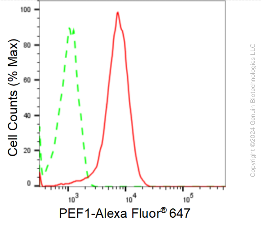
| WB, FC |
|---|---|
| Primary Accession | Q9UBV8 |
| Reactivity | Rat, Human, Mouse |
| Clonality | Monoclonal |
| Isotype | Rabbit IgG |
| Clone Names | 24GB1340 |
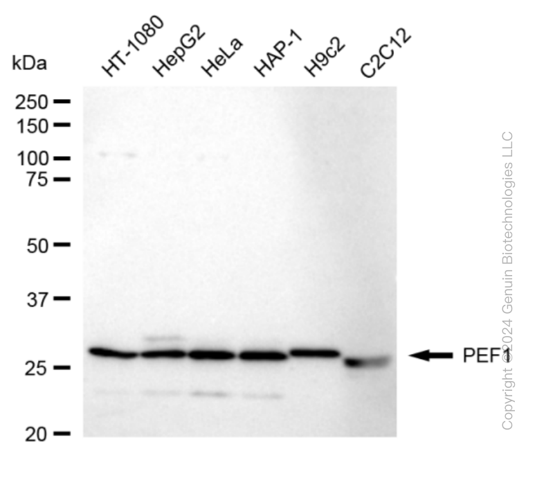
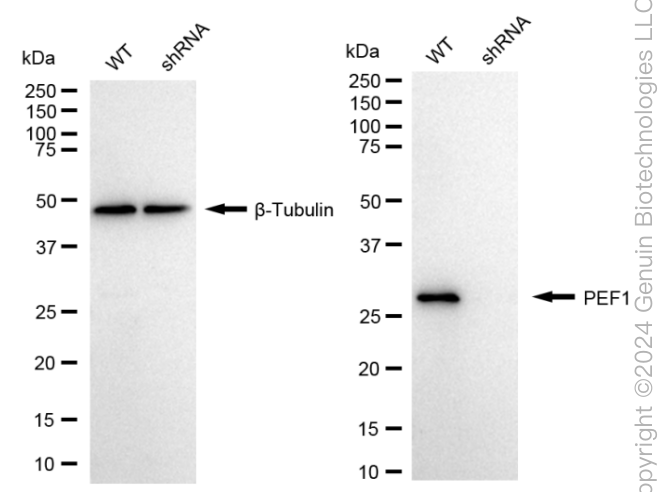
| Calculated MW | Predicted, 30 kDa , observed, 30 kDa |
| Gene Name | PEF1 |
| Aliases | PEF1; Penta-EF-Hand Domain Containing 1; Peflin; PEF1A; PEF Protein With A Long N-Terminal Hydrophobic Domain; ABP32; Epididymis Secretory Sperm Binding Protein; Penta-EF Hand Domain-Containing Protein 1 |
| Immunogen | A synthesized peptide derived from human PEF1 |
| Gene ID | 553115 |
|---|---|
| Other Names | Peflin, PEF protein with a long N-terminal hydrophobic domain, Penta-EF hand domain-containing protein 1 {ECO:0000312|HGNC:HGNC:30009}, PEF1 (HGNC:30009), ABP32 |
| Name | PEF1 (HGNC:30009) |
|---|---|
| Synonyms | ABP32 |
| Function | Calcium-binding protein that acts as an adapter that bridges unrelated proteins or stabilizes weak protein-protein complexes in response to calcium. Together with PDCD6, acts as a calcium-dependent adapter for the BCR(KLHL12) complex, a complex involved in endoplasmic reticulum (ER)-Golgi transport by regulating the size of COPII coats (PubMed:27716508). In response to cytosolic calcium increase, the heterodimer formed with PDCD6 interacts with, and bridges together the BCR(KLHL12) complex and SEC31 (SEC31A or SEC31B), promoting monoubiquitination of SEC31 and subsequent collagen export, which is required for neural crest specification (PubMed:27716508). Its role in the heterodimer formed with PDCD6 is however unclear: some evidence shows that PEF1 and PDCD6 work together and promote association between PDCD6 and SEC31 in presence of calcium (PubMed:27716508). Other reports show that PEF1 dissociates from PDCD6 in presence of calcium, and may act as a negative regulator of PDCD6 (PubMed:11278427). Also acts as a negative regulator of ER-Golgi transport; possibly by inhibiting interaction between PDCD6 and SEC31 (By similarity). |
| Cellular Location | Cytoplasm. Endoplasmic reticulum {ECO:0000250|UniProtKB:Q641Z8}. Membrane; Peripheral membrane protein. Cytoplasmic vesicle, COPII-coated vesicle membrane; Peripheral membrane protein. Note=Membrane-associated in the presence of Ca(2+) (PubMed:11278427). Localizes to endoplasmic reticulum exit site (ERES) (By similarity). {ECO:0000250|UniProtKB:Q641Z8, ECO:0000269|PubMed:11278427} |
Research Areas
Citations (0)

Thousands of laboratories across the world have published research that depended on the performance of antibodies from Abcepta to advance their research. Check out links to articles that cite our products in major peer-reviewed journals, organized by research category.
Submit your citation using an Abcepta antibody to
info@abcepta.com, and receive a free "I Love Antibodies" mug.
info@abcepta.com, and receive a free "I Love Antibodies" mug.

Application Protocols
Provided below are standard protocols that you may find useful for product applications.
Abcepta welcomes feedback from its customers.
If you have used an Abcepta product and would like to share how it has performed, please click on the "Submit Review" button and provide the requested information. Our staff will examine and post your review and contact you if needed.
If you have any additional inquiries please email technical services at tech@abcepta.com.
$ 149.00
$ 499.00
Cat# AGI1674
Ordering Information
United States
AlbaniaAustraliaAustriaBelgiumBosnia & HerzegovinaBrazilBulgariaCanadaCentral AmericaChinaCroatiaCyprusCzech RepublicDenmarkEstoniaFinlandFranceGermanyGreeceHong KongHungaryIcelandIndiaIndonesiaIrelandIsraelItalyJapanLatviaLithuaniaLuxembourgMacedoniaMalaysiaMaltaMexicoNetherlandsNew ZealandNorwayPakistanPolandPortugalRomaniaSerbiaSingaporeSlovakiaSloveniaSouth AfricaSouth KoreaSpainSwedenSwitzerlandTaiwanTurkeyUnited KingdomUnited StatesVietnamWorldwideOthers
USA Headquarters
(888) 735-7227 / (858) 622-0099 or (858) 875-1900
Other Products
Shipping Information
Domestic orders (in stock items)
Shipped out the same day. Orders placed after 1 PM (PST) will ship out the next business day.
International orders
Contact your local distributors
