


 Foundational characteristics of cancer include proliferation, angiogenesis, migration, evasion of apoptosis, and cellular immortality. Find key markers for these cellular processes and antibodies to detect them.
 The SUMOplot™ Analysis Program predicts and scores sumoylation sites in your protein. SUMOylation is a post-translational modification involved in various cellular processes, such as nuclear-cytosolic transport, transcriptional regulation, apoptosis, protein stability, response to stress, and progression through the cell cycle.
 The Autophagy Receptor Motif Plotter predicts and scores autophagy receptor binding sites in your protein. Identifying proteins connected to this pathway is critical to understanding the role of autophagy in physiological as well as pathological processes such as development, differentiation, neurodegenerative diseases, stress, infection, and cancer.
> home > Products > Primary Antibodies > Antibody Collections > KD-Validated Antibodies > KD-Validated Anti-Forkhead box O4 Rabbit Monoclonal Antibody
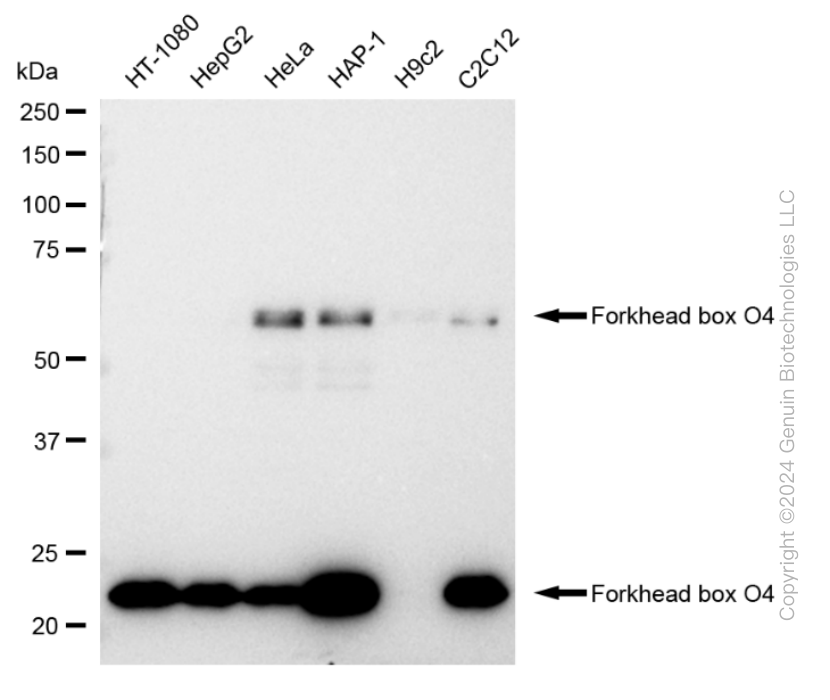
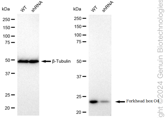
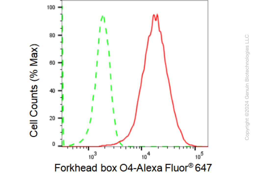
KD-Validated Anti-Forkhead box O4 Rabbit Monoclonal Antibody
Rabbit monoclonal antibody

- SPECIFICATION
- CITATIONS
- PROTOCOLS
- BACKGROUND

Application
| WB, FC, ICC |
|---|---|
| Primary Accession | P98177 |
| Reactivity | Rat, Human, Mouse |
| Clonality | Monoclonal |
| Isotype | Rabbit IgG |
| Clone Names | 23GB1470 |
| Calculated MW | Predicted, 54 kDa , observed, 23,65 kDa |
| Gene Name | FOXO4 |
| Aliases | FOXO4; Forkhead Box O4; AFX1; MLLT7; Myeloid/Lymphoid Or Mixed-Lineage Leukemia (Trithorax Homolog, Drosophila); Translocated To, 7 2 3; Fork Head Domain Transcription Factor AFX1; Forkhead Box Protein O4; AFX; Myeloid/Lymphoid Or Mixed-Lineage Leukemia (Trithorax (Drosophila) Homolog); Translocated To, 7 |
| Immunogen | A synthesized peptide derived from human FoxO4 |
| Gene ID | 4303 |
|---|---|
| Other Names | Forkhead box protein O4, Fork head domain transcription factor AFX1, FOXO4, AFX, AFX1, MLLT7 |
| Name | FOXO4 |
|---|---|
| Synonyms | AFX, AFX1, MLLT7 |
| Function | Transcription factor involved in the regulation of the insulin signaling pathway. Binds to insulin-response elements (IREs) and can activate transcription of IGFBP1. Down-regulates expression of HIF1A and suppresses hypoxia-induced transcriptional activation of HIF1A-modulated genes. Also involved in negative regulation of the cell cycle. Involved in increased proteasome activity in embryonic stem cells (ESCs) by activating expression of PSMD11 in ESCs, leading to enhanced assembly of the 26S proteasome, followed by higher proteasome activity. |
| Cellular Location | Cytoplasm. Nucleus. Note=When phosphorylated, translocated from nucleus to cytoplasm. Dephosphorylation triggers nuclear translocation. Monoubiquitination increases nuclear localization. When deubiquitinated, translocated from nucleus to cytoplasm |
| Tissue Location | Heart, brain, placenta, lung, liver, skeletal muscle, kidney and pancreas. Isoform zeta is most abundant in the liver, kidney, and pancreas |
Research Areas
Citations (0)

Thousands of laboratories across the world have published research that depended on the performance of antibodies from Abcepta to advance their research. Check out links to articles that cite our products in major peer-reviewed journals, organized by research category.
Submit your citation using an Abcepta antibody to
info@abcepta.com, and receive a free "I Love Antibodies" mug.
info@abcepta.com, and receive a free "I Love Antibodies" mug.

Application Protocols
Provided below are standard protocols that you may find useful for product applications.
Abcepta welcomes feedback from its customers.
If you have used an Abcepta product and would like to share how it has performed, please click on the "Submit Review" button and provide the requested information. Our staff will examine and post your review and contact you if needed.
If you have any additional inquiries please email technical services at tech@abcepta.com.
$ 399.20

$ 149.00
Cat# AGI1686
Ordering Information
United States
AlbaniaAustraliaAustriaBelgiumBosnia & HerzegovinaBrazilBulgariaCanadaCentral AmericaChinaCroatiaCyprusCzech RepublicDenmarkEstoniaFinlandFranceGermanyGreeceHong KongHungaryIcelandIndiaIndonesiaIrelandIsraelItalyJapanLatviaLithuaniaLuxembourgMacedoniaMalaysiaMaltaMexicoNetherlandsNew ZealandNorwayPakistanPolandPortugalRomaniaSerbiaSingaporeSlovakiaSloveniaSouth AfricaSouth KoreaSpainSwedenSwitzerlandTaiwanTurkeyUnited KingdomUnited StatesVietnamWorldwideOthers
USA Headquarters
(888) 735-7227 / (858) 622-0099 or (858) 875-1900
Other Products
Shipping Information
Domestic orders (in stock items)
Shipped out the same day. Orders placed after 1 PM (PST) will ship out the next business day.
International orders
Contact your local distributors
