


 Foundational characteristics of cancer include proliferation, angiogenesis, migration, evasion of apoptosis, and cellular immortality. Find key markers for these cellular processes and antibodies to detect them.
 The SUMOplot™ Analysis Program predicts and scores sumoylation sites in your protein. SUMOylation is a post-translational modification involved in various cellular processes, such as nuclear-cytosolic transport, transcriptional regulation, apoptosis, protein stability, response to stress, and progression through the cell cycle.
 The Autophagy Receptor Motif Plotter predicts and scores autophagy receptor binding sites in your protein. Identifying proteins connected to this pathway is critical to understanding the role of autophagy in physiological as well as pathological processes such as development, differentiation, neurodegenerative diseases, stress, infection, and cancer.
> home > Products > Primary Antibodies > Antibody Collections > KD-Validated Antibodies > KD-Validated Anti-VAMP Associated Protein A Rabbit Monoclonal Antibody
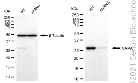
KD-Validated Anti-VAMP Associated Protein A Rabbit Monoclonal Antibody
Rabbit monoclonal antibody
- SPECIFICATION
- CITATIONS
- PROTOCOLS
- BACKGROUND

Application
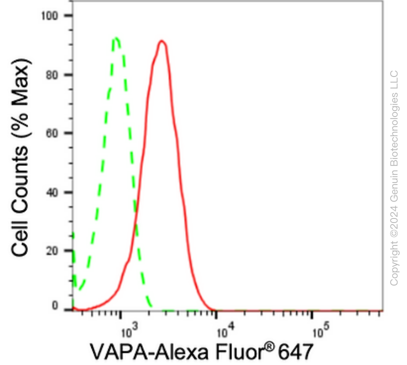
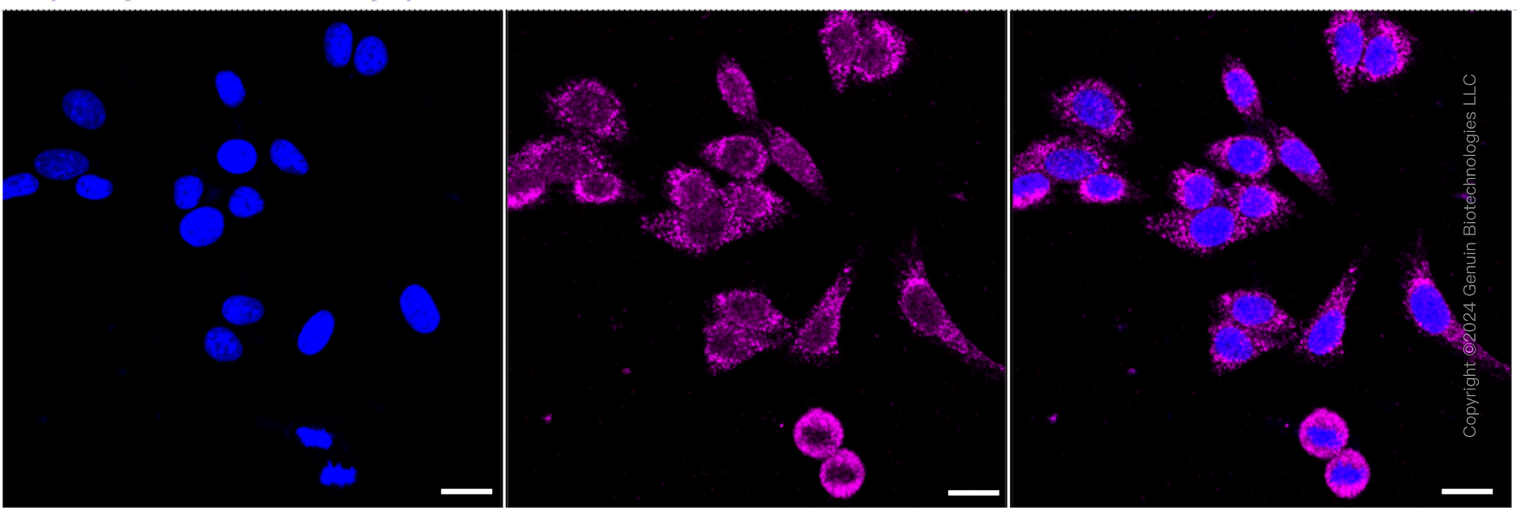
| WB, FC, ICC |
|---|---|
| Primary Accession | Q9P0L0 |
| Reactivity | Rat, Human, Mouse |
| Clonality | Monoclonal |
| Isotype | Rabbit IgG |
| Clone Names | 24GB2580 |
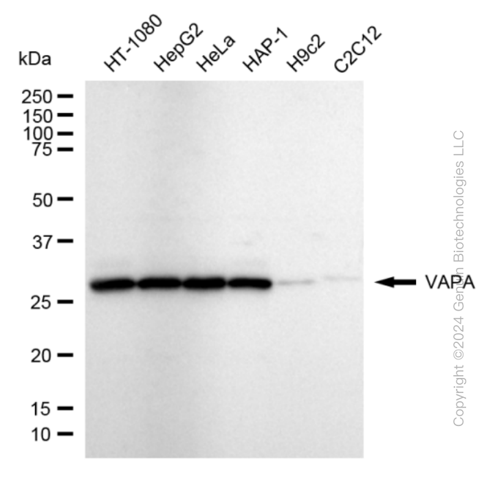
| Calculated MW | Predicted, 28 kDa , observed , 28 kDa |
| Gene Name | VAPA |
| Aliases | VAPA; VAMP Associated Protein A; VAP-A; HVAP-33; VAMP (Vesicle-Associated Membrane Protein)-Associated Protein A, 33kDa; Vesicle-Associated Membrane Protein-Associated Protein A; 33 KDa VAMP-Associated Protein; VAMP-A; VAP-33; VAP33; VAMP (Vesicle-Associated Membrane Protein)-Associated Protein A (33kD); Epididymis Secretory Sperm Binding Protein; VAMP-Associated Protein A |
| Immunogen | A synthesized peptide derived from human VAPA |
| Gene ID | 9218 |
|---|---|
| Other Names | Vesicle-associated membrane protein-associated protein A, VAMP-A, VAMP-associated protein A, VAP-A, 33 kDa VAMP-associated protein {ECO:0000303|Ref.2}, VAP-33, VAPA (HGNC:12648), VAP33 |
| Name | VAPA (HGNC:12648) |
|---|---|
| Synonyms | VAP33 |
| Function | Endoplasmic reticulum (ER)-anchored protein that mediates the formation of contact sites between the ER and endosomes via interaction with FFAT motif-containing proteins such as STARD3 or WDR44 (PubMed:32344433, PubMed:33124732). STARD3-VAPA interaction enables cholesterol transfer from the ER to endosomes (PubMed:33124732). Via interaction with WDR44 participates in neosynthesized protein export (PubMed:32344433). In addition, recruited to the plasma membrane through OSBPL3 binding (PubMed:25447204). The OSBPL3-VAPA complex stimulates RRAS signaling which in turn attenuates integrin beta-1 (ITGB1) activation at the cell surface (PubMed:25447204). With OSBPL3, may regulate ER morphology (PubMed:16143324). May play a role in vesicle trafficking (PubMed:11511104, PubMed:19289470). |
| Cellular Location | Endoplasmic reticulum membrane; Single-pass type IV membrane protein. Cell membrane; Single-pass type IV membrane protein. Cell junction, tight junction. Nucleus membrane {ECO:0000250|UniProtKB:Q9Z270}. Note=Present in the plasma membrane and in intracellular vesicles, together with SNARE proteins. May also associate with the cytoskeleton. Colocalizes with OCLN at the tight junction in polarized epithelial cells. |
| Tissue Location | Ubiquitous. |
Research Areas
Citations (0)

Thousands of laboratories across the world have published research that depended on the performance of antibodies from Abcepta to advance their research. Check out links to articles that cite our products in major peer-reviewed journals, organized by research category.
Submit your citation using an Abcepta antibody to
info@abcepta.com, and receive a free "I Love Antibodies" mug.
info@abcepta.com, and receive a free "I Love Antibodies" mug.

Application Protocols
Provided below are standard protocols that you may find useful for product applications.
Abcepta welcomes feedback from its customers.
If you have used an Abcepta product and would like to share how it has performed, please click on the "Submit Review" button and provide the requested information. Our staff will examine and post your review and contact you if needed.
If you have any additional inquiries please email technical services at tech@abcepta.com.
$ 399.20

$ 149.00
Cat# AGI1766
Ordering Information
United States
AlbaniaAustraliaAustriaBelgiumBosnia & HerzegovinaBrazilBulgariaCanadaCentral AmericaChinaCroatiaCyprusCzech RepublicDenmarkEstoniaFinlandFranceGermanyGreeceHong KongHungaryIcelandIndiaIndonesiaIrelandIsraelItalyJapanLatviaLithuaniaLuxembourgMacedoniaMalaysiaMaltaMexicoNetherlandsNew ZealandNorwayPakistanPolandPortugalRomaniaSerbiaSingaporeSlovakiaSloveniaSouth AfricaSouth KoreaSpainSwedenSwitzerlandTaiwanTurkeyUnited KingdomUnited StatesVietnamWorldwideOthers
USA Headquarters
(888) 735-7227 / (858) 622-0099 or (858) 875-1900
Other Products
Shipping Information
Domestic orders (in stock items)
Shipped out the same day. Orders placed after 1 PM (PST) will ship out the next business day.
International orders
Contact your local distributors
