


 Foundational characteristics of cancer include proliferation, angiogenesis, migration, evasion of apoptosis, and cellular immortality. Find key markers for these cellular processes and antibodies to detect them.
 The SUMOplot™ Analysis Program predicts and scores sumoylation sites in your protein. SUMOylation is a post-translational modification involved in various cellular processes, such as nuclear-cytosolic transport, transcriptional regulation, apoptosis, protein stability, response to stress, and progression through the cell cycle.
 The Autophagy Receptor Motif Plotter predicts and scores autophagy receptor binding sites in your protein. Identifying proteins connected to this pathway is critical to understanding the role of autophagy in physiological as well as pathological processes such as development, differentiation, neurodegenerative diseases, stress, infection, and cancer.
> home > Products > Primary Antibodies > Antibody Collections > KD-Validated Antibodies > KD-Validated Anti-Prolyl 4-Hydroxylase Subunit Beta Mouse Monoclonal Antibody
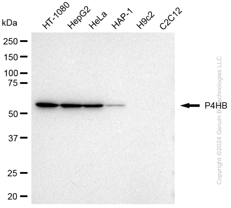
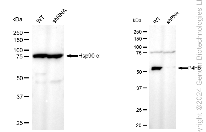
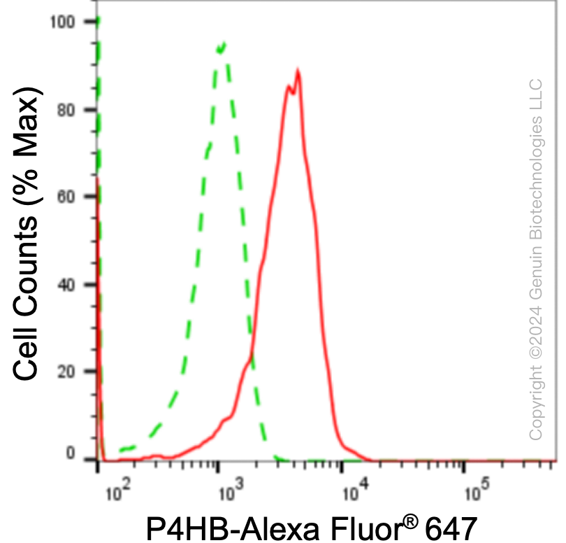
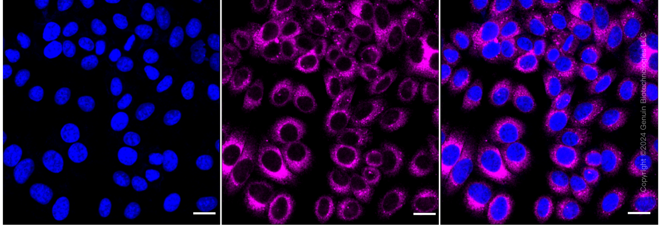
KD-Validated Anti-Prolyl 4-Hydroxylase Subunit Beta Mouse Monoclonal Antibody
Mouse monoclonal antibody
- SPECIFICATION
- CITATIONS
- PROTOCOLS
- BACKGROUND

Application
| WB, FC, ICC |
|---|---|
| Primary Accession | P07237 |
| Reactivity | Human |
| Clonality | Monoclonal |
| Isotype | Mouse IgG1 kappa |
| Clone Names | 24GB2780 |
| Calculated MW | Predicted, 57 kDa , o bserved , 57 kDa |
| Gene Name | P4HB |
| Aliases | P4HB; Prolyl 4-Hydroxylase Subunit Beta; PDIA1; PDI; P4Hbeta; ERBA2L; PROHB; PO4HB; PO4DB; DSI; GIT; Procollagen-Proline, 2-Oxoglutarate 4-Dioxygenase (Proline 4-Hydroxylase), Beta Polypeptide; Protein Disulfide Isomerase Family A, Member 1; Protein Disulfide Isomerase-Associated 1; Cellular Thyroid Hormone-Binding Protein; Prolyl 4-Hydroxylase, Beta Polypeptide; Collagen Prolyl 4-Hydroxylase Beta; Protein Disulfide-Isomerase; EC 5.3.4.1; P55; Procollagen-Proline, 2-Oxoglutarate 4-Dioxygenase (Proline 4-Hydroxylase), Beta Polypeptide (Protein Disulfide Isomerase; Thyroid Hormone Binding Protein P55) ; Procollagen-Proline, 2-Oxoglutarate 4-Dioxygenase (Proline 4-Hydroxylase), Beta Polypeptide (Protein Disulfide Isomerase-Associated 1); Protein Disulfide Isomerase/Oxidoreductase; Glutathione-Insulin Transhydrogenase; Thyroid Hormone-Binding Protein P55; Testicular Secretory Protein Li 32; Protocollagen Hydroxylase; CLCRP1; PHDB |
| Immunogen | Recombinant protein of human PDI |
| Gene ID | 5034 |
|---|---|
| Other Names | Protein disulfide-isomerase, PDI, 5.3.4.1, Cellular thyroid hormone-binding protein, Prolyl 4-hydroxylase subunit beta, p55, P4HB, ERBA2L, PDI, PDIA1, PO4DB |
| Name | P4HB |
|---|---|
| Synonyms | ERBA2L, PDI, PDIA1, PO4DB |
| Function | This multifunctional protein catalyzes the formation, breakage and rearrangement of disulfide bonds. At the cell surface, seems to act as a reductase that cleaves disulfide bonds of proteins attached to the cell. May therefore cause structural modifications of exofacial proteins. Inside the cell, seems to form/rearrange disulfide bonds of nascent proteins. At high concentrations and following phosphorylation by FAM20C, functions as a chaperone that inhibits aggregation of misfolded proteins (PubMed:32149426). At low concentrations, facilitates aggregation (anti-chaperone activity). May be involved with other chaperones in the structural modification of the TG precursor in hormone biogenesis. Also acts as a structural subunit of various enzymes such as prolyl 4-hydroxylase and microsomal triacylglycerol transfer protein MTTP. Receptor for LGALS9; the interaction retains P4HB at the cell surface of Th2 T helper cells, increasing disulfide reductase activity at the plasma membrane, altering the plasma membrane redox state and enhancing cell migration (PubMed:21670307). |
| Cellular Location | Endoplasmic reticulum. Endoplasmic reticulum lumen. Melanosome. Cell membrane; Peripheral membrane protein. Note=Highly abundant. In some cell types, seems to be also secreted or associated with the plasma membrane, where it undergoes constant shedding and replacement from intracellular sources (Probable). Localizes near CD4-enriched regions on lymphoid cell surfaces (PubMed:11181151). Identified by mass spectrometry in melanosome fractions from stage I to stage IV (PubMed:10636893) Colocalizes with MTTP in the endoplasmic reticulum (PubMed:23475612) {ECO:0000269|PubMed:10636893, ECO:0000269|PubMed:11181151, ECO:0000269|PubMed:23475612, ECO:0000305} |
Research Areas
Citations (0)

Thousands of laboratories across the world have published research that depended on the performance of antibodies from Abcepta to advance their research. Check out links to articles that cite our products in major peer-reviewed journals, organized by research category.
Submit your citation using an Abcepta antibody to
info@abcepta.com, and receive a free "I Love Antibodies" mug.
info@abcepta.com, and receive a free "I Love Antibodies" mug.

Application Protocols
Provided below are standard protocols that you may find useful for product applications.
Abcepta welcomes feedback from its customers.
If you have used an Abcepta product and would like to share how it has performed, please click on the "Submit Review" button and provide the requested information. Our staff will examine and post your review and contact you if needed.
If you have any additional inquiries please email technical services at tech@abcepta.com.
$ 399.20

$ 149.00
Cat# AGI1780
Ordering Information
United States
AlbaniaAustraliaAustriaBelgiumBosnia & HerzegovinaBrazilBulgariaCanadaCentral AmericaChinaCroatiaCyprusCzech RepublicDenmarkEstoniaFinlandFranceGermanyGreeceHong KongHungaryIcelandIndiaIndonesiaIrelandIsraelItalyJapanLatviaLithuaniaLuxembourgMacedoniaMalaysiaMaltaMexicoNetherlandsNew ZealandNorwayPakistanPolandPortugalRomaniaSerbiaSingaporeSlovakiaSloveniaSouth AfricaSouth KoreaSpainSwedenSwitzerlandTaiwanTurkeyUnited KingdomUnited StatesVietnamWorldwideOthers
USA Headquarters
(888) 735-7227 / (858) 622-0099 or (858) 875-1900
Other Products
Shipping Information
Domestic orders (in stock items)
Shipped out the same day. Orders placed after 1 PM (PST) will ship out the next business day.
International orders
Contact your local distributors
