


 Foundational characteristics of cancer include proliferation, angiogenesis, migration, evasion of apoptosis, and cellular immortality. Find key markers for these cellular processes and antibodies to detect them.
 The SUMOplot™ Analysis Program predicts and scores sumoylation sites in your protein. SUMOylation is a post-translational modification involved in various cellular processes, such as nuclear-cytosolic transport, transcriptional regulation, apoptosis, protein stability, response to stress, and progression through the cell cycle.
 The Autophagy Receptor Motif Plotter predicts and scores autophagy receptor binding sites in your protein. Identifying proteins connected to this pathway is critical to understanding the role of autophagy in physiological as well as pathological processes such as development, differentiation, neurodegenerative diseases, stress, infection, and cancer.
> home > Products > Primary Antibodies > Antibody Collections > KD-Validated Antibodies > KD-Validated Anti-MITF Mouse Monoclonal Antibody
KD-Validated Anti-MITF Mouse Monoclonal Antibody
Mouse monoclonal antibody
- SPECIFICATION
- CITATIONS
- PROTOCOLS
- BACKGROUND

Application
| WB, ICC |
|---|---|
| Primary Accession | O75030 |
| Reactivity | Human |
| Clonality | Monoclonal |
| Isotype | Mouse IgG1 kappa |
| Clone Names | 24GB2905 |
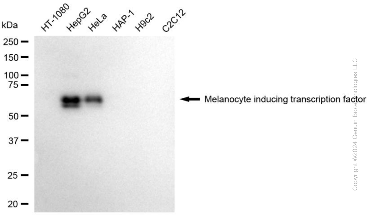
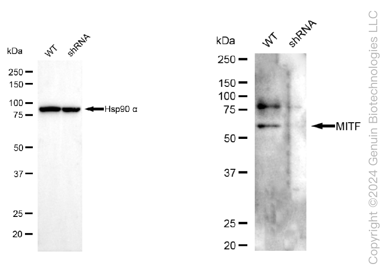
| Calculated MW | Predicted, 59 kDa, observed, 57 kDa |
| Gene Name | MITF |
| Aliases | MITF; Melanocyte Inducing Transcription Factor; BHLHe32; Microphthalmia-Associated Transcription Factor; MI; Melanogenesis Associated Transcription Factor; Class E Basic Helix-Loop-Helix Protein 32; WS2A; WS2; Microphtalmia-Associated Transcription Factor; Homolog Of Mouse Microphthalmia; Waardenburg Syndrome, Type 2A; BHLHE32; COMMAD; MITF-A; CMM8 |
| Immunogen | Recombinant protein of human MITF |
| Gene ID | 4286 |
|---|---|
| Other Names | Microphthalmia-associated transcription factor, Class E basic helix-loop-helix protein 32, bHLHe32, MITF {ECO:0000303|PubMed:8069297, ECO:0000312|HGNC:HGNC:7105} |
| Name | MITF {ECO:0000303|PubMed:8069297, ECO:0000312|HGNC:HGNC:7105} |
|---|---|
| Function | Transcription factor that acts as a master regulator of melanocyte survival and differentiation as well as melanosome biogenesis (PubMed:10587587, PubMed:22647378, PubMed:27889061, PubMed:9647758). Binds to M-boxes (5'-TCATGTG-3') and symmetrical DNA sequences (E-boxes) (5'-CACGTG-3') found in the promoter of pigmentation genes, such as tyrosinase (TYR) (PubMed:10587587, PubMed:22647378, PubMed:27889061, PubMed:9647758). Involved in the cellular response to amino acid availability by acting downstream of MTOR: in the presence of nutrients, MITF phosphorylation by MTOR promotes its inactivation (PubMed:36608670). Upon starvation or lysosomal stress, inhibition of MTOR induces MITF dephosphorylation, resulting in transcription factor activity (PubMed:36608670). Plays an important role in melanocyte development by regulating the expression of tyrosinase (TYR) and tyrosinase-related protein 1 (TYRP1) (PubMed:10587587, PubMed:22647378, PubMed:27889061, PubMed:9647758). Plays a critical role in the differentiation of various cell types, such as neural crest-derived melanocytes, mast cells, osteoclasts and optic cup-derived retinal pigment epithelium (PubMed:10587587, PubMed:22647378, PubMed:27889061, PubMed:9647758). |
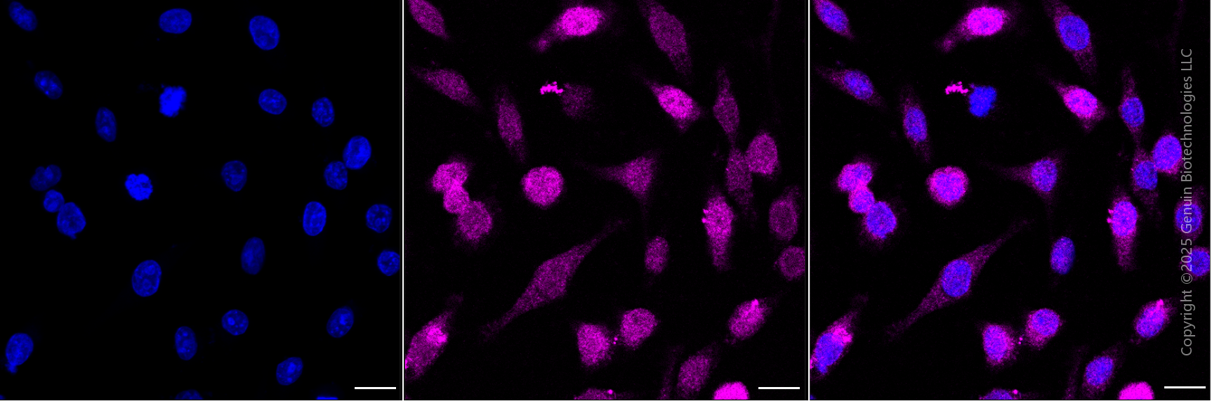
| Cellular Location | Nucleus. Cytoplasm. Lysosome membrane Note=When nutrients are present, recruited to the lysosomal membrane via association with GDP-bound RagC/RRAGC (or RagD/RRAGD): it is then phosphorylated by MTOR (PubMed:23401004, PubMed:36608670) Phosphorylation by MTOR promotes ubiquitination and degradation (PubMed:36608670). Conversely, inhibition of mTORC1, starvation and lysosomal disruption, promotes dephosphorylation and translocation to the nucleus (PubMed:36608670). Phosphorylation by MARK3/cTAK1 promotes association with 14-3-3/YWHA adapters and retention in the cytosol (PubMed:16822840). |
| Tissue Location | Expressed in melanocytes (at protein level). [Isoform C2]: Expressed in the kidney and retinal pigment epithelium. [Isoform H2]: Expressed in the kidney. [Isoform Mdel]: Expressed in melanocytes. |
Research Areas
Citations (0)

Thousands of laboratories across the world have published research that depended on the performance of antibodies from Abcepta to advance their research. Check out links to articles that cite our products in major peer-reviewed journals, organized by research category.
Submit your citation using an Abcepta antibody to
info@abcepta.com, and receive a free "I Love Antibodies" mug.
info@abcepta.com, and receive a free "I Love Antibodies" mug.

Application Protocols
Provided below are standard protocols that you may find useful for product applications.
Abcepta welcomes feedback from its customers.
If you have used an Abcepta product and would like to share how it has performed, please click on the "Submit Review" button and provide the requested information. Our staff will examine and post your review and contact you if needed.
If you have any additional inquiries please email technical services at tech@abcepta.com.
$ 399.20

$ 149.00
Cat# AGI1787
Ordering Information
United States
AlbaniaAustraliaAustriaBelgiumBosnia & HerzegovinaBrazilBulgariaCanadaCentral AmericaChinaCroatiaCyprusCzech RepublicDenmarkEstoniaFinlandFranceGermanyGreeceHong KongHungaryIcelandIndiaIndonesiaIrelandIsraelItalyJapanLatviaLithuaniaLuxembourgMacedoniaMalaysiaMaltaMexicoNetherlandsNew ZealandNorwayPakistanPolandPortugalRomaniaSerbiaSingaporeSlovakiaSloveniaSouth AfricaSouth KoreaSpainSwedenSwitzerlandTaiwanTurkeyUnited KingdomUnited StatesVietnamWorldwideOthers
USA Headquarters
(888) 735-7227 / (858) 622-0099 or (858) 875-1900
Other Products
Shipping Information
Domestic orders (in stock items)
Shipped out the same day. Orders placed after 1 PM (PST) will ship out the next business day.
International orders
Contact your local distributors
