


 Foundational characteristics of cancer include proliferation, angiogenesis, migration, evasion of apoptosis, and cellular immortality. Find key markers for these cellular processes and antibodies to detect them.
 The SUMOplot™ Analysis Program predicts and scores sumoylation sites in your protein. SUMOylation is a post-translational modification involved in various cellular processes, such as nuclear-cytosolic transport, transcriptional regulation, apoptosis, protein stability, response to stress, and progression through the cell cycle.
 The Autophagy Receptor Motif Plotter predicts and scores autophagy receptor binding sites in your protein. Identifying proteins connected to this pathway is critical to understanding the role of autophagy in physiological as well as pathological processes such as development, differentiation, neurodegenerative diseases, stress, infection, and cancer.
> home > Products > Primary Antibodies > Antibody Collections > KD-Validated Antibodies > KD-Validated Anti-Mitogen-Activated Protein Kinase 1 Rabbit Monoclonal Antibody
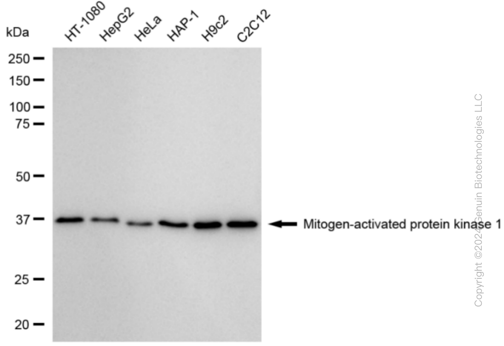
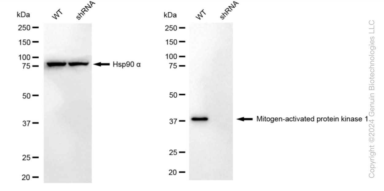
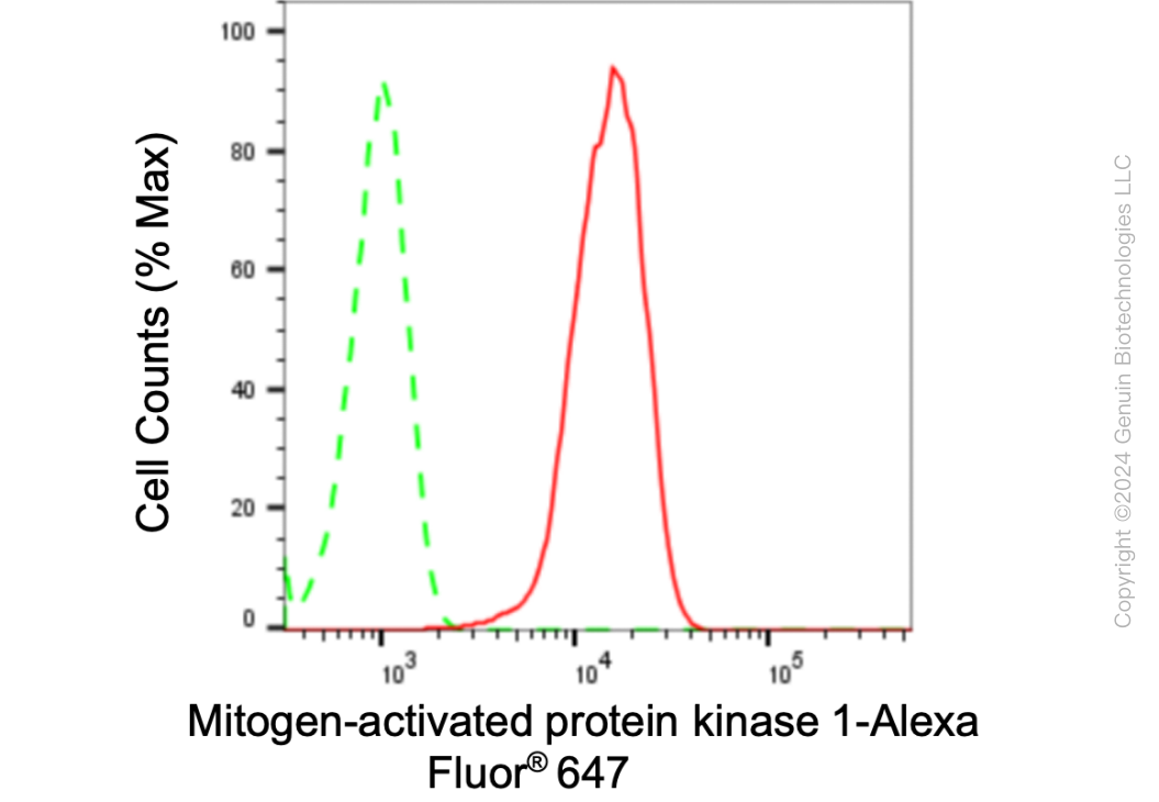
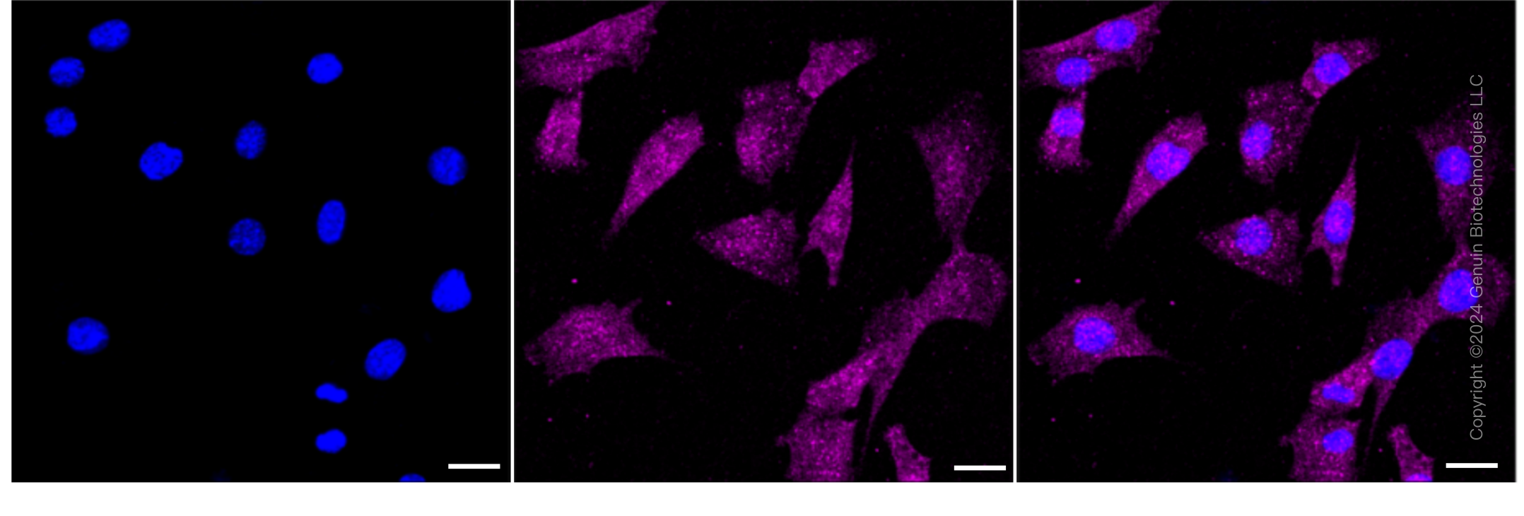
KD-Validated Anti-Mitogen-Activated Protein Kinase 1 Rabbit Monoclonal Antibody
Rabbit monoclonal antibody
- SPECIFICATION
- CITATIONS
- PROTOCOLS
- BACKGROUND

Application
| WB, FC, ICC |
|---|---|
| Primary Accession | P28482 |
| Reactivity | Rat, Human, Mouse |
| Clonality | Monoclonal |
| Isotype | Rabbit IgG |
| Clone Names | 24GB3095 |
| Calculated MW | Predicted, 41 kDa, observed, 41 kDa |
| Gene Name | MAPK1 |
| Aliases | MAPK1; Mitogen-Activated Protein Kinase 1; ERK2; Extracellular Signal Regulated Kinase 2; P41mapk; MAPK; PRKM1; PRKM2; ERK; Mitogen-Activated Protein Kinase 2; MAP Kinase 1; MAP Kinase 2; EC 2.7.11.24; P42-MAPK; MAPK 2; ERK-2; ERT1; Protein Tyrosine Kinase ERK2; MAP Kinase Isoform P42; EC 2.7.11; P42MAPK; MAPK 1; NS13; P38; P40; P41 |
| Immunogen | A synthesized peptide derived from human ERK2 |
| Gene ID | 5594 |
|---|---|
| Other Names | Mitogen-activated protein kinase 1, MAP kinase 1, MAPK 1, 2.7.11.24, ERT1, Extracellular signal-regulated kinase 2, ERK-2, MAP kinase isoform p42, p42-MAPK, Mitogen-activated protein kinase 2, MAP kinase 2, MAPK 2, MAPK1 (HGNC:6871), ERK2, PRKM1, PRKM2 |
| Name | MAPK1 (HGNC:6871) |
|---|---|
| Synonyms | ERK2, PRKM1, PRKM2 |
| Function | Serine/threonine kinase which acts as an essential component of the MAP kinase signal transduction pathway. MAPK1/ERK2 and MAPK3/ERK1 are the 2 MAPKs which play an important role in the MAPK/ERK cascade. They participate also in a signaling cascade initiated by activated KIT and KITLG/SCF. Depending on the cellular context, the MAPK/ERK cascade mediates diverse biological functions such as cell growth, adhesion, survival and differentiation through the regulation of transcription, translation, cytoskeletal rearrangements. The MAPK/ERK cascade also plays a role in initiation and regulation of meiosis, mitosis, and postmitotic functions in differentiated cells by phosphorylating a number of transcription factors. About 160 substrates have already been discovered for ERKs. Many of these substrates are localized in the nucleus, and seem to participate in the regulation of transcription upon stimulation. However, other substrates are found in the cytosol as well as in other cellular organelles, and those are responsible for processes such as translation, mitosis and apoptosis. Moreover, the MAPK/ERK cascade is also involved in the regulation of the endosomal dynamics, including lysosome processing and endosome cycling through the perinuclear recycling compartment (PNRC); as well as in the fragmentation of the Golgi apparatus during mitosis. The substrates include transcription factors (such as ATF2, BCL6, ELK1, ERF, FOS, HSF4 or SPZ1), cytoskeletal elements (such as CANX, CTTN, GJA1, MAP2, MAPT, PXN, SORBS3 or STMN1), regulators of apoptosis (such as BAD, BTG2, CASP9, DAPK1, IER3, MCL1 or PPARG), regulators of translation (such as EIF4EBP1 and FXR1) and a variety of other signaling-related molecules (like ARHGEF2, DCC, FRS2 or GRB10). Protein kinases (such as RAF1, RPS6KA1/RSK1, RPS6KA3/RSK2, RPS6KA2/RSK3, RPS6KA6/RSK4, SYK, MKNK1/MNK1, MKNK2/MNK2, RPS6KA5/MSK1, RPS6KA4/MSK2, MAPKAPK3 or MAPKAPK5) and phosphatases (such as DUSP1, DUSP4, DUSP6 or DUSP16) are other substrates which enable the propagation the MAPK/ERK signal to additional cytosolic and nuclear targets, thereby extending the specificity of the cascade. Mediates phosphorylation of TPR in response to EGF stimulation. May play a role in the spindle assembly checkpoint. Phosphorylates PML and promotes its interaction with PIN1, leading to PML degradation. Phosphorylates CDK2AP2 (By similarity). Phosphorylates phosphoglycerate kinase PGK1 under hypoxic conditions to promote its targeting to the mitochondrion and suppress the formation of acetyl-coenzyme A from pyruvate (PubMed:26942675). |
| Cellular Location | Cytoplasm, cytoskeleton, spindle. Nucleus. Cytoplasm, cytoskeleton, microtubule organizing center, centrosome. Cytoplasm. Membrane, caveola {ECO:0000250|UniProtKB:P63086}. Cell junction, focal adhesion {ECO:0000250|UniProtKB:P63085}. Note=Associated with the spindle during prometaphase and metaphase (By similarity). PEA15-binding and phosphorylated DAPK1 promote its cytoplasmic retention. Phosphorylation at Ser- 246 and Ser-248 as well as autophosphorylation at Thr-190 promote nuclear localization. |
Research Areas
Citations (0)

Thousands of laboratories across the world have published research that depended on the performance of antibodies from Abcepta to advance their research. Check out links to articles that cite our products in major peer-reviewed journals, organized by research category.
Submit your citation using an Abcepta antibody to
info@abcepta.com, and receive a free "I Love Antibodies" mug.
info@abcepta.com, and receive a free "I Love Antibodies" mug.

Application Protocols
Provided below are standard protocols that you may find useful for product applications.
Abcepta welcomes feedback from its customers.
If you have used an Abcepta product and would like to share how it has performed, please click on the "Submit Review" button and provide the requested information. Our staff will examine and post your review and contact you if needed.
If you have any additional inquiries please email technical services at tech@abcepta.com.
$ 399.20

$ 149.00
Cat# AGI1792
Ordering Information
United States
AlbaniaAustraliaAustriaBelgiumBosnia & HerzegovinaBrazilBulgariaCanadaCentral AmericaChinaCroatiaCyprusCzech RepublicDenmarkEstoniaFinlandFranceGermanyGreeceHong KongHungaryIcelandIndiaIndonesiaIrelandIsraelItalyJapanLatviaLithuaniaLuxembourgMacedoniaMalaysiaMaltaMexicoNetherlandsNew ZealandNorwayPakistanPolandPortugalRomaniaSerbiaSingaporeSlovakiaSloveniaSouth AfricaSouth KoreaSpainSwedenSwitzerlandTaiwanTurkeyUnited KingdomUnited StatesVietnamWorldwideOthers
USA Headquarters
(888) 735-7227 / (858) 622-0099 or (858) 875-1900
Other Products
Shipping Information
Domestic orders (in stock items)
Shipped out the same day. Orders placed after 1 PM (PST) will ship out the next business day.
International orders
Contact your local distributors
