


 Foundational characteristics of cancer include proliferation, angiogenesis, migration, evasion of apoptosis, and cellular immortality. Find key markers for these cellular processes and antibodies to detect them.
 The SUMOplot™ Analysis Program predicts and scores sumoylation sites in your protein. SUMOylation is a post-translational modification involved in various cellular processes, such as nuclear-cytosolic transport, transcriptional regulation, apoptosis, protein stability, response to stress, and progression through the cell cycle.
 The Autophagy Receptor Motif Plotter predicts and scores autophagy receptor binding sites in your protein. Identifying proteins connected to this pathway is critical to understanding the role of autophagy in physiological as well as pathological processes such as development, differentiation, neurodegenerative diseases, stress, infection, and cancer.
> home > Products > Primary Antibodies > Antibody Collections > KD-Validated Antibodies > KD-Validated Anti-CD46 Rabbit Monoclonal Antibody
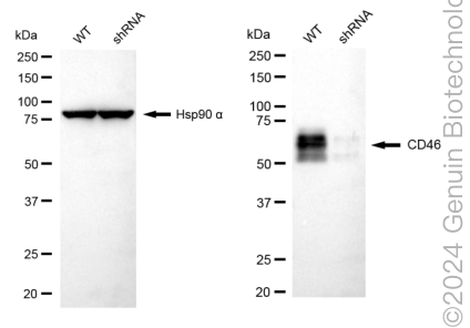
KD-Validated Anti-CD46 Rabbit Monoclonal Antibody
Rabbit monoclonal antibody
- SPECIFICATION
- CITATIONS
- PROTOCOLS
- BACKGROUND

Application
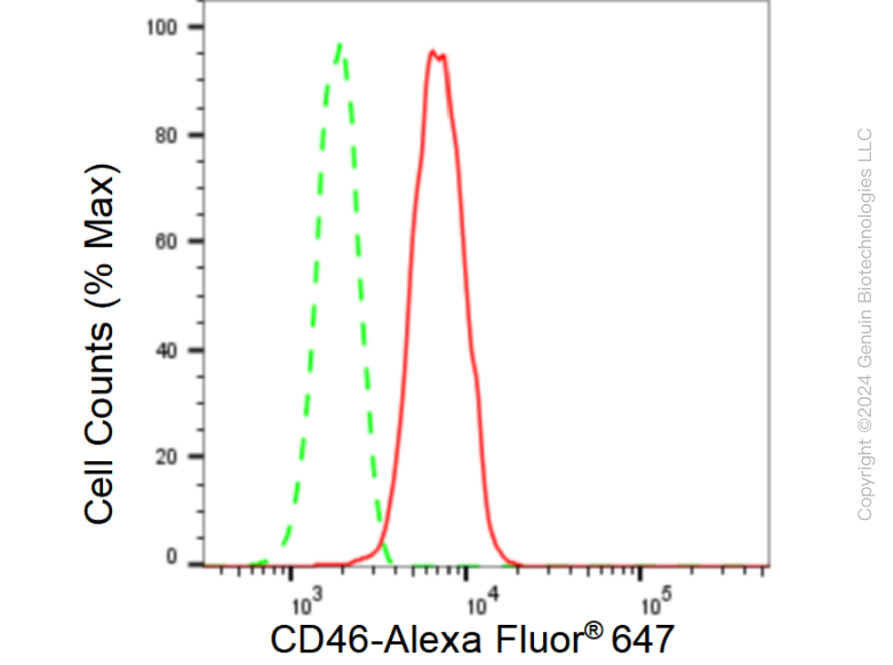
| WB, FC |
|---|---|
| Primary Accession | P15529 |
| Reactivity | Human |
| Clonality | Monoclonal |
| Isotype | Rabbit IgG |
| Clone Names | 23GB5720 |
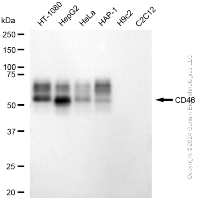
| Calculated MW | Predicted, 44 kDa , observed , 50-70 kDa |
| Gene Name | CD46 |
| Aliases | CD46; CD46 Molecule; TLX; TRA2.10; MIC10; MCP; Membrane Cofactor Protein (CD46, Trophoblast-Lymphocyte Cross-Reactive Antigen); Antigen Identified By Monoclonal Antibody TRA-2-10; Trophoblast-Lymphocyte Cross-Reactive Antigen; CD46 Molecule, Complement Regulatory Protein; CD46 Antigen, Complement Regulatory Protein; Trophoblast Leukocyte Common Antigen; Membrane Cofactor Protein; MGC26544; Complement Membrane Cofactor Protein; Trophoblast Leucocyte Common Antigen; Measles Virus Receptor; CD46 Antigen; AHUS2 |
| Immunogen | A synthesized peptide derived from human CD46 |
| Gene ID | 4179 |
|---|---|
| Other Names | Membrane cofactor protein, TLX, Trophoblast leukocyte common antigen, CD46, CD46, MCP, MIC10 |
| Name | CD46 |
|---|---|
| Synonyms | MCP, MIC10 |
| Function | Acts as a cofactor for complement factor I, a serine protease which protects autologous cells against complement-mediated injury by cleaving C3b and C4b deposited on host tissue. May be involved in the fusion of the spermatozoa with the oocyte during fertilization. Also acts as a costimulatory factor for T-cells which induces the differentiation of CD4+ into T-regulatory 1 cells. T-regulatory 1 cells suppress immune responses by secreting interleukin-10, and therefore are thought to prevent autoimmunity. |
| Cellular Location | Cytoplasmic vesicle, secretory vesicle, acrosome inner membrane; Single-pass type I membrane protein. Note=Inner acrosomal membrane of spermatozoa. Internalized upon binding of Measles virus, Herpesvirus 6 or Neisseria gonorrhoeae, which results in an increased susceptibility of infected cells to complement-mediated injury. In cancer cells or cells infected by Neisseria, shedding leads to a soluble peptide |
| Tissue Location | Expressed by all cells except erythrocytes. |
Research Areas
Citations (0)

Thousands of laboratories across the world have published research that depended on the performance of antibodies from Abcepta to advance their research. Check out links to articles that cite our products in major peer-reviewed journals, organized by research category.
Submit your citation using an Abcepta antibody to
info@abcepta.com, and receive a free "I Love Antibodies" mug.
info@abcepta.com, and receive a free "I Love Antibodies" mug.

Application Protocols
Provided below are standard protocols that you may find useful for product applications.
Abcepta welcomes feedback from its customers.
If you have used an Abcepta product and would like to share how it has performed, please click on the "Submit Review" button and provide the requested information. Our staff will examine and post your review and contact you if needed.
If you have any additional inquiries please email technical services at tech@abcepta.com.
$ 399.20

$ 149.00
Cat# AGI1794
Ordering Information
United States
AlbaniaAustraliaAustriaBelgiumBosnia & HerzegovinaBrazilBulgariaCanadaCentral AmericaChinaCroatiaCyprusCzech RepublicDenmarkEstoniaFinlandFranceGermanyGreeceHong KongHungaryIcelandIndiaIndonesiaIrelandIsraelItalyJapanLatviaLithuaniaLuxembourgMacedoniaMalaysiaMaltaMexicoNetherlandsNew ZealandNorwayPakistanPolandPortugalRomaniaSerbiaSingaporeSlovakiaSloveniaSouth AfricaSouth KoreaSpainSwedenSwitzerlandTaiwanTurkeyUnited KingdomUnited StatesVietnamWorldwideOthers
USA Headquarters
(888) 735-7227 / (858) 622-0099 or (858) 875-1900
Other Products
Shipping Information
Domestic orders (in stock items)
Shipped out the same day. Orders placed after 1 PM (PST) will ship out the next business day.
International orders
Contact your local distributors
