


 Foundational characteristics of cancer include proliferation, angiogenesis, migration, evasion of apoptosis, and cellular immortality. Find key markers for these cellular processes and antibodies to detect them.
 The SUMOplot™ Analysis Program predicts and scores sumoylation sites in your protein. SUMOylation is a post-translational modification involved in various cellular processes, such as nuclear-cytosolic transport, transcriptional regulation, apoptosis, protein stability, response to stress, and progression through the cell cycle.
 The Autophagy Receptor Motif Plotter predicts and scores autophagy receptor binding sites in your protein. Identifying proteins connected to this pathway is critical to understanding the role of autophagy in physiological as well as pathological processes such as development, differentiation, neurodegenerative diseases, stress, infection, and cancer.
> home > Products > Primary Antibodies > Antibody Collections > Prostate negative > KD-Validated Anti-Fibroblast Growth Factor 2 Rabbit Monoclonal Antibody
KD-Validated Anti-Fibroblast Growth Factor 2 Rabbit Monoclonal Antibody
Rabbit monoclonal antibody
- SPECIFICATION
- CITATIONS
- PROTOCOLS
- BACKGROUND

Application
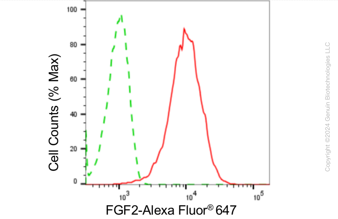
| WB, FC |
|---|---|
| Primary Accession | P09038 |
| Reactivity | Human |
| Clonality | Monoclonal |
| Isotype | Rabbit IgG |
| Clone Names | 24GB4420 |
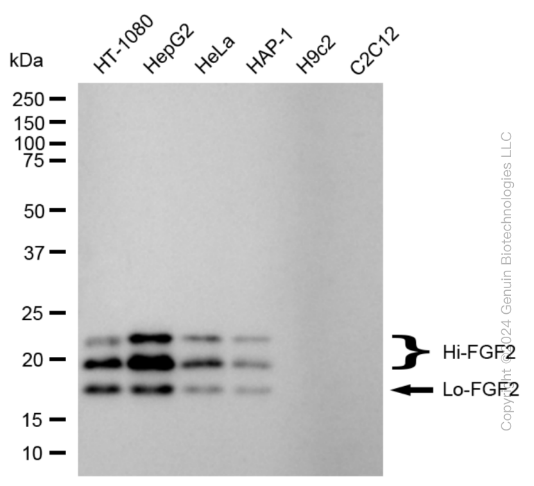
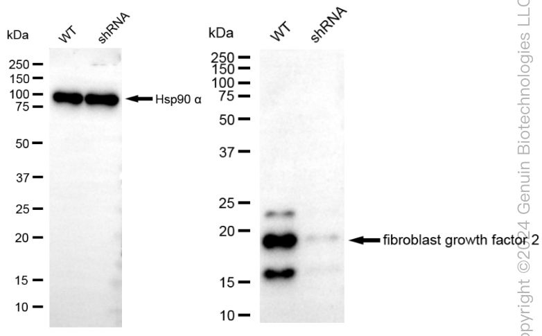
| Calculated MW | Predicted, 31 kDa, observed, 18-24kDa |
| Gene Name | FGF2 |
| Aliases | FGF2; Fibroblast Growth Factor 2; FGFB; Fibroblast Growth Factor 2 (Basic); Heparin-Binding Growth Factor 2; HBGF-2; FGF-2; BFGF; Basic Fibroblast Growth Factor BFGF; Basic Fibroblast Growth Factor; Prostatropin |
| Immunogen | Recombinant protein of human FGF2 |
| Gene ID | 2247 |
|---|---|
| Other Names | Fibroblast growth factor 2, FGF-2, Basic fibroblast growth factor, bFGF, Heparin-binding growth factor 2, HBGF-2, FGF2, FGFB |
| Name | FGF2 |
|---|---|
| Synonyms | FGFB |
| Function | Acts as a ligand for FGFR1, FGFR2, FGFR3 and FGFR4 (PubMed:8663044). Also acts as an integrin ligand which is required for FGF2 signaling (PubMed:28302677). Binds to integrin ITGAV:ITGB3 (PubMed:28302677). Plays an important role in the regulation of cell survival, cell division, cell differentiation and cell migration (PubMed:28302677, PubMed:8663044). Functions as a potent mitogen in vitro (PubMed:1721615, PubMed:3732516, PubMed:3964259). Can induce angiogenesis (PubMed:23469107, PubMed:28302677). Mediates phosphorylation of ERK1/2 and thereby promotes retinal lens fiber differentiation (PubMed:29501879). |
| Cellular Location | Secreted. Nucleus. Note=Exported from cells by an endoplasmic reticulum (ER)/Golgi-independent mechanism. Unconventional secretion of FGF2 occurs by direct translocation across the plasma membrane (PubMed:20230531). Binding of exogenous FGF2 to FGFR facilitates endocytosis followed by translocation of FGF2 across endosomal membrane into the cytosol (PubMed:22321063). Nuclear import from the cytosol requires the classical nuclear import machinery, involving proteins KPNA1 and KPNB1, as well as CEP57 (PubMed:22321063) |
| Tissue Location | Expressed in granulosa and cumulus cells. Expressed in hepatocellular carcinoma cells, but not in non-cancerous liver tissue. |
Research Areas
Citations (0)

Thousands of laboratories across the world have published research that depended on the performance of antibodies from Abcepta to advance their research. Check out links to articles that cite our products in major peer-reviewed journals, organized by research category.
Submit your citation using an Abcepta antibody to
info@abcepta.com, and receive a free "I Love Antibodies" mug.
info@abcepta.com, and receive a free "I Love Antibodies" mug.

Application Protocols
Provided below are standard protocols that you may find useful for product applications.
Abcepta welcomes feedback from its customers.
If you have used an Abcepta product and would like to share how it has performed, please click on the "Submit Review" button and provide the requested information. Our staff will examine and post your review and contact you if needed.
If you have any additional inquiries please email technical services at tech@abcepta.com.
$ 399.20

$ 149.00
Cat# AGI1817
Ordering Information
United States
AlbaniaAustraliaAustriaBelgiumBosnia & HerzegovinaBrazilBulgariaCanadaCentral AmericaChinaCroatiaCyprusCzech RepublicDenmarkEstoniaFinlandFranceGermanyGreeceHong KongHungaryIcelandIndiaIndonesiaIrelandIsraelItalyJapanLatviaLithuaniaLuxembourgMacedoniaMalaysiaMaltaMexicoNetherlandsNew ZealandNorwayPakistanPolandPortugalRomaniaSerbiaSingaporeSlovakiaSloveniaSouth AfricaSouth KoreaSpainSwedenSwitzerlandTaiwanTurkeyUnited KingdomUnited StatesVietnamWorldwideOthers
USA Headquarters
(888) 735-7227 / (858) 622-0099 or (858) 875-1900
Other Products
Shipping Information
Domestic orders (in stock items)
Shipped out the same day. Orders placed after 1 PM (PST) will ship out the next business day.
International orders
Contact your local distributors
