


 Foundational characteristics of cancer include proliferation, angiogenesis, migration, evasion of apoptosis, and cellular immortality. Find key markers for these cellular processes and antibodies to detect them.
 The SUMOplot™ Analysis Program predicts and scores sumoylation sites in your protein. SUMOylation is a post-translational modification involved in various cellular processes, such as nuclear-cytosolic transport, transcriptional regulation, apoptosis, protein stability, response to stress, and progression through the cell cycle.
 The Autophagy Receptor Motif Plotter predicts and scores autophagy receptor binding sites in your protein. Identifying proteins connected to this pathway is critical to understanding the role of autophagy in physiological as well as pathological processes such as development, differentiation, neurodegenerative diseases, stress, infection, and cancer.
> home > Products > Primary Antibodies > Antibody Collections > KD-Validated Antibodies > KD-Validated Anti-HDAC3 Rabbit Monoclonal Antibody
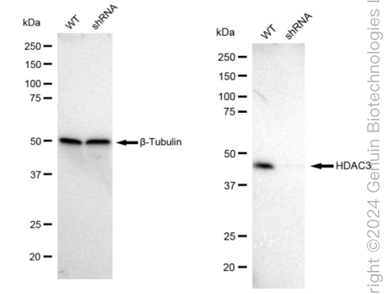
KD-Validated Anti-HDAC3 Rabbit Monoclonal Antibody
Rabbit monoclonal antibody
- SPECIFICATION
- CITATIONS
- PROTOCOLS
- BACKGROUND

Application
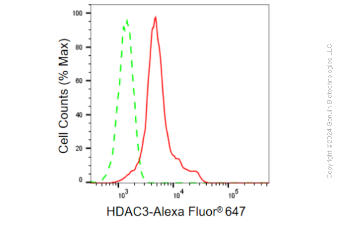
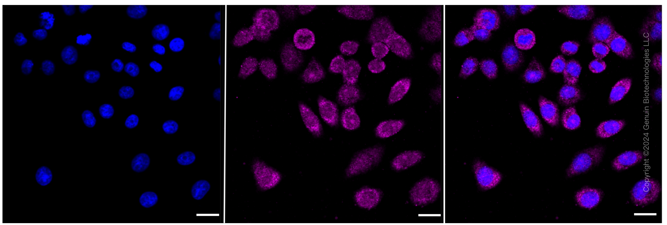
| WB, FC, ICC |
|---|---|
| Primary Accession | O15379 |
| Reactivity | Rat, Human, Mouse |
| Clonality | Monoclonal |
| Isotype | Rabbit IgG |
| Clone Names | 23GB860 |
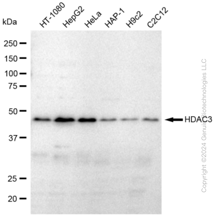
| Calculated MW | Predicted, 49 kDa , observed, 49 kDa |
| Gene Name | HDAC3 |
| Aliases | Histone Deacetylase 3; RPD3-2; HD3 KDAC3; RPD3; Protein Deacetylase HDAC3; Protein Deacylase HDAC3; EC 3.5.1.98; SMAP45; EC 3.5.1.- |
| Immunogen | A synthesized peptide derived from human HDAC3 |
| Gene ID | 8841 |
|---|---|
| Other Names | Histone deacetylase 3, HD3, 3.5.1.98, Protein deacetylase HDAC3, 3.5.1.-, Protein deacylase HDAC3, 3.5.1.-, RPD3-2, SMAP45, HDAC3 |
| Name | HDAC3 |
|---|---|
| Function | Histone deacetylase that catalyzes the deacetylation of lysine residues on the N-terminal part of the core histones (H2A, H2B, H3 and H4), and some other non-histone substrates (PubMed:21030595, PubMed:21444723, PubMed:23911289, PubMed:25301942, PubMed:28167758, PubMed:28497810, PubMed:32404892, PubMed:22230954). Histone deacetylation gives a tag for epigenetic repression and plays an important role in transcriptional regulation, cell cycle progression and developmental events (PubMed:23911289). Histone deacetylases act via the formation of large multiprotein complexes, such as N-Cor repressor complex, which activate the histone deacetylase activity (PubMed:23911289, PubMed:22230954). Participates in the BCL6 transcriptional repressor activity by deacetylating the H3 'Lys-27' (H3K27) on enhancer elements, antagonizing EP300 acetyltransferase activity and repressing proximal gene expression (PubMed:23911289). Acts as a molecular chaperone for shuttling phosphorylated NR2C1 to PML bodies for sumoylation (By similarity). Contributes, together with XBP1 isoform 1, to the activation of NFE2L2-mediated HMOX1 transcription factor gene expression in a PI(3)K/mTORC2/Akt-dependent signaling pathway leading to endothelial cell (EC) survival under disturbed flow/oxidative stress (PubMed:25190803). Regulates both the transcriptional activation and repression phases of the circadian clock in a deacetylase activity-independent manner (By similarity). During the activation phase, promotes the accumulation of ubiquitinated BMAL1 at the E-boxes and during the repression phase, blocks FBXL3-mediated CRY1/2 ubiquitination and promotes the interaction of CRY1 and BMAL1 (By similarity). The NCOR1-HDAC3 complex regulates the circadian expression of the core clock gene BMAL1 and the genes involved in lipid metabolism in the liver (By similarity). Also functions as a deacetylase for non-histone targets, such as KAT5, MEF2D, MAPK14, RARA and STAT3 (PubMed:15653507, PubMed:21030595, PubMed:21444723, PubMed:25301942, PubMed:28167758). Serves as a corepressor of RARA, mediating its deacetylation and repression, leading to inhibition of RARE DNA element binding (PubMed:28167758). In association with RARA, plays a role in the repression of microRNA-10a and thereby in the inflammatory response (PubMed:28167758). In addition to protein deacetylase activity, also acts as a protein-lysine deacylase by recognizing other acyl groups: catalyzes removal of (2E)-butenoyl (crotonyl), lactoyl (lactyl) and 2-hydroxyisobutanoyl (2- hydroxyisobutyryl) acyl groups from lysine residues, leading to protein decrotonylation, delactylation and de-2-hydroxyisobutyrylation, respectively (PubMed:28497810, PubMed:29192674, PubMed:34608293, PubMed:35044827). Catalyzes decrotonylation of MAPRE1/EB1 (PubMed:34608293). Mediates delactylation NBN/NBS1, thereby inhibiting DNA double-strand breaks (DSBs) via homologous recombination (HR) (PubMed:38961290). |
| Cellular Location | Nucleus. Chromosome. Cytoplasm. Cytoplasm, cytosol. Note=Colocalizes with XBP1 and AKT1 in the cytoplasm (PubMed:25190803). Predominantly expressed in the nucleus in the presence of CCAR2 (PubMed:21030595) |
| Tissue Location | Widely expressed.. |
Research Areas
Citations (0)

Thousands of laboratories across the world have published research that depended on the performance of antibodies from Abcepta to advance their research. Check out links to articles that cite our products in major peer-reviewed journals, organized by research category.
Submit your citation using an Abcepta antibody to
info@abcepta.com, and receive a free "I Love Antibodies" mug.
info@abcepta.com, and receive a free "I Love Antibodies" mug.

Application Protocols
Provided below are standard protocols that you may find useful for product applications.
Abcepta welcomes feedback from its customers.
If you have used an Abcepta product and would like to share how it has performed, please click on the "Submit Review" button and provide the requested information. Our staff will examine and post your review and contact you if needed.
If you have any additional inquiries please email technical services at tech@abcepta.com.
$ 399.20

$ 149.00
Cat# AGI1847
Ordering Information
United States
AlbaniaAustraliaAustriaBelgiumBosnia & HerzegovinaBrazilBulgariaCanadaCentral AmericaChinaCroatiaCyprusCzech RepublicDenmarkEstoniaFinlandFranceGermanyGreeceHong KongHungaryIcelandIndiaIndonesiaIrelandIsraelItalyJapanLatviaLithuaniaLuxembourgMacedoniaMalaysiaMaltaMexicoNetherlandsNew ZealandNorwayPakistanPolandPortugalRomaniaSerbiaSingaporeSlovakiaSloveniaSouth AfricaSouth KoreaSpainSwedenSwitzerlandTaiwanTurkeyUnited KingdomUnited StatesVietnamWorldwideOthers
USA Headquarters
(888) 735-7227 / (858) 622-0099 or (858) 875-1900
Other Products
Shipping Information
Domestic orders (in stock items)
Shipped out the same day. Orders placed after 1 PM (PST) will ship out the next business day.
International orders
Contact your local distributors
