


 Foundational characteristics of cancer include proliferation, angiogenesis, migration, evasion of apoptosis, and cellular immortality. Find key markers for these cellular processes and antibodies to detect them.
 The SUMOplot™ Analysis Program predicts and scores sumoylation sites in your protein. SUMOylation is a post-translational modification involved in various cellular processes, such as nuclear-cytosolic transport, transcriptional regulation, apoptosis, protein stability, response to stress, and progression through the cell cycle.
 The Autophagy Receptor Motif Plotter predicts and scores autophagy receptor binding sites in your protein. Identifying proteins connected to this pathway is critical to understanding the role of autophagy in physiological as well as pathological processes such as development, differentiation, neurodegenerative diseases, stress, infection, and cancer.
> home > Products > Primary Antibodies > Antibody Collections > KD-Validated Antibodies > KD-Validated Anti-Cofilin 1 Rabbit Monoclonal Antibody
KD-Validated Anti-Cofilin 1 Rabbit Monoclonal Antibody
Rabbit monoclonal antibody
- SPECIFICATION
- CITATIONS
- PROTOCOLS
- BACKGROUND

Application
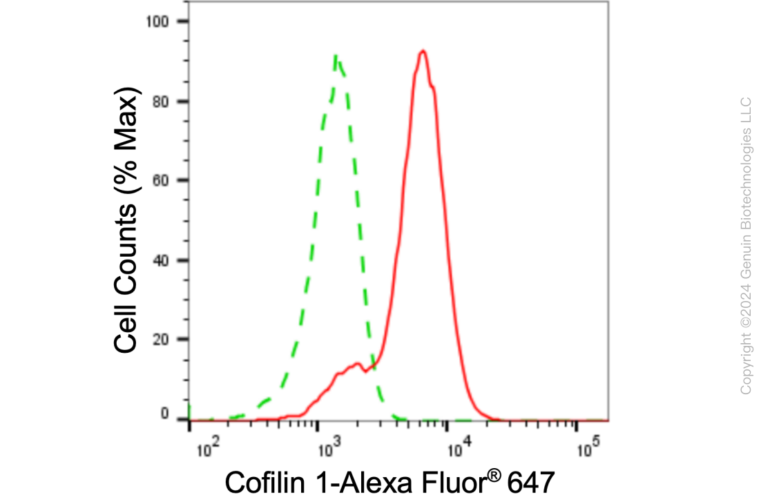
| WB, FC |
|---|---|
| Primary Accession | P23528 |
| Reactivity | Rat, Human, Mouse |
| Clonality | Monoclonal |
| Isotype | Rabbit IgG |
| Clone Names | 24GB4775 |
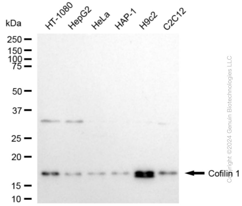
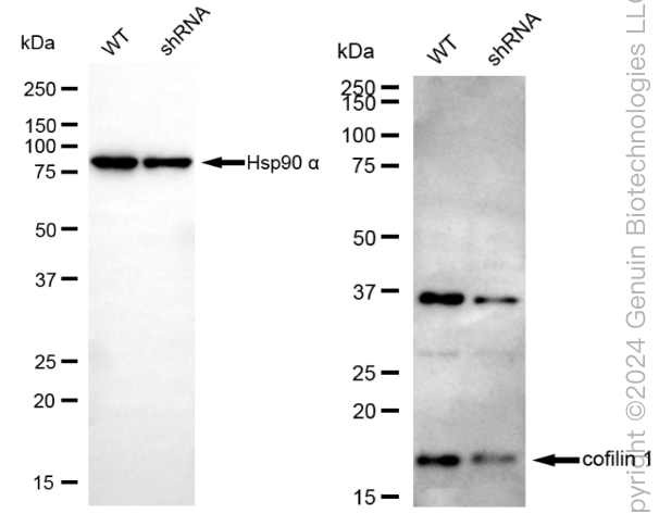
| Calculated MW | Predicted, 19 kDa,observed, 19 kDa |
| Gene Name | CFL1 |
| Aliases | CFL1; Cofilin 1; CFL; Cofilin 1 (Non-Muscle); 18 KDa Phosphoprotein; Cofilin-1; P18; Epididymis Secretory Protein Li 15; Cofilin, Non-Muscle Isoform; HEL-S-15; Cofilin |
| Immunogen | A synthesized peptide derived from human Cofilin |
| Gene ID | 1072 |
|---|---|
| Other Names | Cofilin-1, 18 kDa phosphoprotein, p18, Cofilin, non-muscle isoform, CFL1, CFL |
| Name | CFL1 |
|---|---|
| Synonyms | CFL |
| Function | Binds to F-actin and exhibits pH-sensitive F-actin depolymerizing activity (PubMed:11812157). In conjunction with the subcortical maternal complex (SCMC), plays an essential role for zygotes to progress beyond the first embryonic cell divisions via regulation of actin dynamics (PubMed:15580268). Required for the centralization of the mitotic spindle and symmetric division of zygotes (By similarity). Plays a role in the regulation of cell morphology and cytoskeletal organization in epithelial cells (PubMed:21834987). Required for the up-regulation of atypical chemokine receptor ACKR2 from endosomal compartment to cell membrane, increasing its efficiency in chemokine uptake and degradation (PubMed:23633677). Required for neural tube morphogenesis and neural crest cell migration (By similarity). |
| Cellular Location | Nucleus matrix. Cytoplasm, cytoskeleton. Cell projection, ruffle membrane; Peripheral membrane protein; Cytoplasmic side. Cell projection, lamellipodium membrane; Peripheral membrane protein; Cytoplasmic side. Cell projection, lamellipodium {ECO:0000250|UniProtKB:P18760}. Cell projection, growth cone {ECO:0000250|UniProtKB:P18760}. Cell projection, axon {ECO:0000250|UniProtKB:P18760}. Note=Colocalizes with the actin cytoskeleton in membrane ruffles and lamellipodia. Detected at the cleavage furrow and contractile ring during cytokinesis. Almost completely in nucleus in cells exposed to heat shock or 10% dimethyl sulfoxide |
| Tissue Location | Widely distributed in various tissues. |
Research Areas
Citations (0)

Thousands of laboratories across the world have published research that depended on the performance of antibodies from Abcepta to advance their research. Check out links to articles that cite our products in major peer-reviewed journals, organized by research category.
Submit your citation using an Abcepta antibody to
info@abcepta.com, and receive a free "I Love Antibodies" mug.
info@abcepta.com, and receive a free "I Love Antibodies" mug.

Application Protocols
Provided below are standard protocols that you may find useful for product applications.
Abcepta welcomes feedback from its customers.
If you have used an Abcepta product and would like to share how it has performed, please click on the "Submit Review" button and provide the requested information. Our staff will examine and post your review and contact you if needed.
If you have any additional inquiries please email technical services at tech@abcepta.com.
$ 399.20

$ 149.00
Cat# AGI1858
Ordering Information
United States
AlbaniaAustraliaAustriaBelgiumBosnia & HerzegovinaBrazilBulgariaCanadaCentral AmericaChinaCroatiaCyprusCzech RepublicDenmarkEstoniaFinlandFranceGermanyGreeceHong KongHungaryIcelandIndiaIndonesiaIrelandIsraelItalyJapanLatviaLithuaniaLuxembourgMacedoniaMalaysiaMaltaMexicoNetherlandsNew ZealandNorwayPakistanPolandPortugalRomaniaSerbiaSingaporeSlovakiaSloveniaSouth AfricaSouth KoreaSpainSwedenSwitzerlandTaiwanTurkeyUnited KingdomUnited StatesVietnamWorldwideOthers
USA Headquarters
(888) 735-7227 / (858) 622-0099 or (858) 875-1900
Other Products
Shipping Information
Domestic orders (in stock items)
Shipped out the same day. Orders placed after 1 PM (PST) will ship out the next business day.
International orders
Contact your local distributors
