


 Foundational characteristics of cancer include proliferation, angiogenesis, migration, evasion of apoptosis, and cellular immortality. Find key markers for these cellular processes and antibodies to detect them.
 The SUMOplot™ Analysis Program predicts and scores sumoylation sites in your protein. SUMOylation is a post-translational modification involved in various cellular processes, such as nuclear-cytosolic transport, transcriptional regulation, apoptosis, protein stability, response to stress, and progression through the cell cycle.
 The Autophagy Receptor Motif Plotter predicts and scores autophagy receptor binding sites in your protein. Identifying proteins connected to this pathway is critical to understanding the role of autophagy in physiological as well as pathological processes such as development, differentiation, neurodegenerative diseases, stress, infection, and cancer.
> home > Products > Primary Antibodies > Antibody Collections > KD-Validated Antibodies > KD-Validated Anti-BCR Rabbit Monoclonal Antibody
KD-Validated Anti-BCR Rabbit Monoclonal Antibody
Rabbit monoclonal antibody

- SPECIFICATION
- CITATIONS
- PROTOCOLS
- BACKGROUND

Application
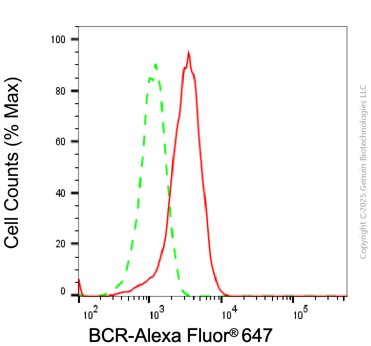
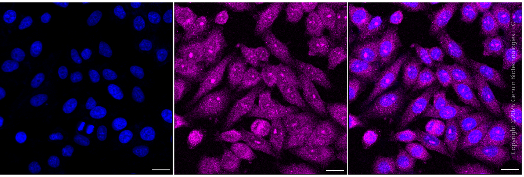
| WB, FC, ICC |
|---|---|
| Primary Accession | P11274 |
| Reactivity | Human, Mouse |
| Clonality | Monoclonal |
| Isotype | Rabbit IgG |
| Clone Names | 24GB6945 |
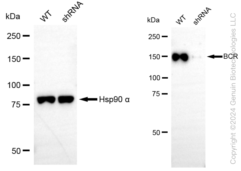
| Calculated MW | Predicted, 143 kDa, observed, 160 kDa |
| Gene Name | BCR |
| Aliases | BCR; BCR Activator Of RhoGEF And GTPase; D22S662; D22S11; BCR1; CML; PHL; ALL; BCR, RhoGEF And GTPase Activating Protein; Breakpoint Cluster Region Protein; Renal Carcinoma Antigen NY-REN-26; Breakpoint Cluster Region; EC 2.7.11.1; BCR/FGFR1 Chimera Protein; FGFR1/BCR Chimera Protein |
| Immunogen | A synthesized peptide derived from human Bcr |
| Gene ID | 613 |
|---|---|
| Other Names | Breakpoint cluster region protein, 2.7.11.1, Renal carcinoma antigen NY-REN-26, BCR (HGNC:1014), BCR1, D22S11 |
| Name | BCR (HGNC:1014) |
|---|---|
| Synonyms | BCR1, D22S11 |
| Function | Protein with a unique structure having two opposing regulatory activities toward small GTP-binding proteins. The C-terminus is a GTPase-activating protein (GAP) domain which stimulates GTP hydrolysis by RAC1, RAC2 and CDC42. Accelerates the intrinsic rate of GTP hydrolysis of RAC1 or CDC42, leading to down-regulation of the active GTP-bound form (PubMed:17116687, PubMed:1903516, PubMed:7479768). The central Dbl homology (DH) domain functions as guanine nucleotide exchange factor (GEF) that modulates the GTPases CDC42, RHOA and RAC1. Promotes the conversion of CDC42, RHOA and RAC1 from the GDP-bound to the GTP-bound form (PubMed:23940119, PubMed:7479768). The amino terminus contains an intrinsic kinase activity (PubMed:1657398). Functions as an important negative regulator of neuronal RAC1 activity (By similarity). Regulates macrophage functions such as CSF1-directed motility and phagocytosis through the modulation of RAC1 activity (PubMed:17116687). Plays a major role as a RHOA GEF in keratinocytes being involved in focal adhesion formation and keratinocyte differentiation (PubMed:23940119). |
| Cellular Location | Postsynaptic density {ECO:0000250|UniProtKB:Q6PAJ1}. Cell projection, dendritic spine {ECO:0000250|UniProtKB:Q6PAJ1}. Cell projection, axon {ECO:0000250|UniProtKB:Q6PAJ1}. Synapse {ECO:0000250|UniProtKB:F1LXF1} |
Research Areas
Citations (0)

Thousands of laboratories across the world have published research that depended on the performance of antibodies from Abcepta to advance their research. Check out links to articles that cite our products in major peer-reviewed journals, organized by research category.
Submit your citation using an Abcepta antibody to
info@abcepta.com, and receive a free "I Love Antibodies" mug.
info@abcepta.com, and receive a free "I Love Antibodies" mug.

Application Protocols
Provided below are standard protocols that you may find useful for product applications.
Abcepta welcomes feedback from its customers.
If you have used an Abcepta product and would like to share how it has performed, please click on the "Submit Review" button and provide the requested information. Our staff will examine and post your review and contact you if needed.
If you have any additional inquiries please email technical services at tech@abcepta.com.
$ 399.20

$ 149.00
Cat# AGI1983
Ordering Information
United States
AlbaniaAustraliaAustriaBelgiumBosnia & HerzegovinaBrazilBulgariaCanadaCentral AmericaChinaCroatiaCyprusCzech RepublicDenmarkEstoniaFinlandFranceGermanyGreeceHong KongHungaryIcelandIndiaIndonesiaIrelandIsraelItalyJapanLatviaLithuaniaLuxembourgMacedoniaMalaysiaMaltaMexicoNetherlandsNew ZealandNorwayPakistanPolandPortugalRomaniaSerbiaSingaporeSlovakiaSloveniaSouth AfricaSouth KoreaSpainSwedenSwitzerlandTaiwanTurkeyUnited KingdomUnited StatesVietnamWorldwideOthers
USA Headquarters
(888) 735-7227 / (858) 622-0099 or (858) 875-1900
Shipping Information
Domestic orders (in stock items)
Shipped out the same day. Orders placed after 1 PM (PST) will ship out the next business day.
International orders
Contact your local distributors
