


 Foundational characteristics of cancer include proliferation, angiogenesis, migration, evasion of apoptosis, and cellular immortality. Find key markers for these cellular processes and antibodies to detect them.
 The SUMOplot™ Analysis Program predicts and scores sumoylation sites in your protein. SUMOylation is a post-translational modification involved in various cellular processes, such as nuclear-cytosolic transport, transcriptional regulation, apoptosis, protein stability, response to stress, and progression through the cell cycle.
 The Autophagy Receptor Motif Plotter predicts and scores autophagy receptor binding sites in your protein. Identifying proteins connected to this pathway is critical to understanding the role of autophagy in physiological as well as pathological processes such as development, differentiation, neurodegenerative diseases, stress, infection, and cancer.
> home > Products > Primary Antibodies > Antibody Collections > KD-Validated Antibodies > KD-Validated Anti-ATP5C1 Mouse Monoclonal Antibody
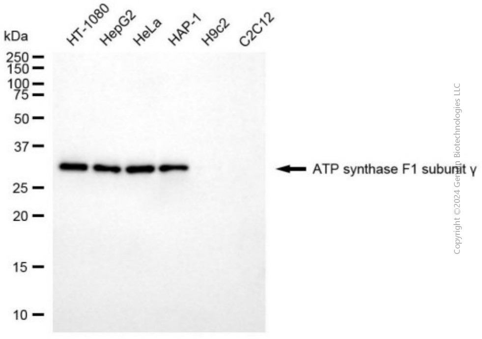
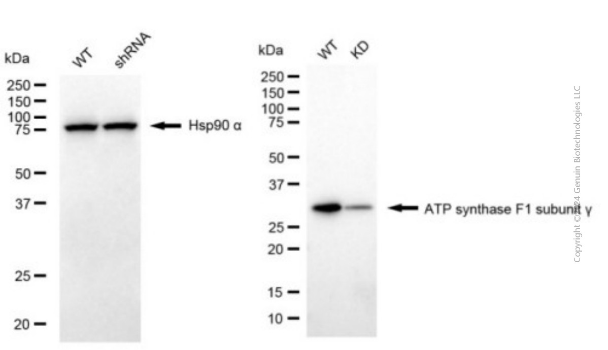
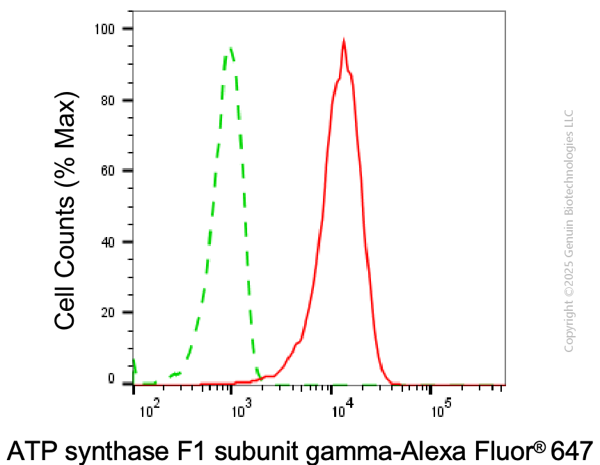
KD-Validated Anti-ATP5C1 Mouse Monoclonal Antibody
Mouse monoclonal antibody
- SPECIFICATION
- CITATIONS
- PROTOCOLS
- BACKGROUND

Application
| WB, FC |
|---|---|
| Primary Accession | P36542 |
| Reactivity | Human |
| Clonality | Monoclonal |
| Isotype | Mouse IgG1 |
| Clone Names | 24GB7990 |
| Calculated MW | Predicted, 33 kDa, observed, 33 kDa |
| Gene Name | ATP5F1C |
| Aliases | ATP5F1C; ATP Synthase F1 Subunit Gamma; ATP5CL1; ATP5C1; ATP5C; ATP Synthase, H+ Transporting, Mitochondrial F1 Complex, Gamma Polypeptide 1; ATP Synthase Subunit Gamma, Mitochondrial; F-ATPase Gamma Subunit; Mitochondrial ATP Synthase, Gamma Subunit 1; ATP Synthase Gamma Chain, Mitochondrial |
| Immunogen | Recombinant protein of human ATP5C1 |
| Gene ID | 509 |
|---|---|
| Other Names | ATP synthase F(1) complex subunit gamma, mitochondrial, ATP synthase F1 subunit gamma {ECO:0000312|HGNC:HGNC:833}, F-ATPase gamma subunit, ATP5F1C (HGNC:833) |
| Name | ATP5F1C (HGNC:833) |
|---|---|
| Function | Subunit gamma, of the mitochondrial membrane ATP synthase complex (F(1)F(0) ATP synthase or Complex V) that produces ATP from ADP in the presence of a proton gradient across the membrane which is generated by electron transport complexes of the respiratory chain (PubMed:37244256). ATP synthase complex consist of a soluble F(1) head domain - the catalytic core - and a membrane F(1) domain - the membrane proton channel (PubMed:37244256). These two domains are linked by a central stalk rotating inside the F(1) region and a stationary peripheral stalk (PubMed:37244256). During catalysis, ATP synthesis in the catalytic domain of F(1) is coupled via a rotary mechanism of the central stalk subunits to proton translocation (Probable). In vivo, can only synthesize ATP although its ATP hydrolase activity can be activated artificially in vitro (By similarity). With the central stalk subunit delta, is essential for the biogenesis of F(1) catalytic part of the ATP synthase complex namely in the formation of F1 assembly intermediate (PubMed:29499186). |
| Cellular Location | Mitochondrion inner membrane {ECO:0000250|UniProtKB:P05631}; Peripheral membrane protein {ECO:0000250|UniProtKB:P05631}; Matrix side {ECO:0000250|UniProtKB:P05631} |
| Tissue Location | Isoform Heart is expressed specifically in the heart and skeletal muscle, which require rapid energy supply. Isoform Liver is expressed in the brain, liver and kidney. Isoform Heart and Isoform Liver are expressed in the skin, intestine, stomach and aorta |
Research Areas
Citations (0)

Thousands of laboratories across the world have published research that depended on the performance of antibodies from Abcepta to advance their research. Check out links to articles that cite our products in major peer-reviewed journals, organized by research category.
Submit your citation using an Abcepta antibody to
info@abcepta.com, and receive a free "I Love Antibodies" mug.
info@abcepta.com, and receive a free "I Love Antibodies" mug.

Application Protocols
Provided below are standard protocols that you may find useful for product applications.
Abcepta welcomes feedback from its customers.
If you have used an Abcepta product and would like to share how it has performed, please click on the "Submit Review" button and provide the requested information. Our staff will examine and post your review and contact you if needed.
If you have any additional inquiries please email technical services at tech@abcepta.com.
$ 399.20

$ 149.00
Cat# AGI2016
Ordering Information
United States
AlbaniaAustraliaAustriaBelgiumBosnia & HerzegovinaBrazilBulgariaCanadaCentral AmericaChinaCroatiaCyprusCzech RepublicDenmarkEstoniaFinlandFranceGermanyGreeceHong KongHungaryIcelandIndiaIndonesiaIrelandIsraelItalyJapanLatviaLithuaniaLuxembourgMacedoniaMalaysiaMaltaMexicoNetherlandsNew ZealandNorwayPakistanPolandPortugalRomaniaSerbiaSingaporeSlovakiaSloveniaSouth AfricaSouth KoreaSpainSwedenSwitzerlandTaiwanTurkeyUnited KingdomUnited StatesVietnamWorldwideOthers
USA Headquarters
(888) 735-7227 / (858) 622-0099 or (858) 875-1900
Other Products
Shipping Information
Domestic orders (in stock items)
Shipped out the same day. Orders placed after 1 PM (PST) will ship out the next business day.
International orders
Contact your local distributors
