


 Foundational characteristics of cancer include proliferation, angiogenesis, migration, evasion of apoptosis, and cellular immortality. Find key markers for these cellular processes and antibodies to detect them.
 The SUMOplot™ Analysis Program predicts and scores sumoylation sites in your protein. SUMOylation is a post-translational modification involved in various cellular processes, such as nuclear-cytosolic transport, transcriptional regulation, apoptosis, protein stability, response to stress, and progression through the cell cycle.
 The Autophagy Receptor Motif Plotter predicts and scores autophagy receptor binding sites in your protein. Identifying proteins connected to this pathway is critical to understanding the role of autophagy in physiological as well as pathological processes such as development, differentiation, neurodegenerative diseases, stress, infection, and cancer.
> home > Products > Primary Antibodies > Antibody Collections > KD-Validated Antibodies > KD-Validated Anti-HERPUD1 Mouse Monoclonal Antibody
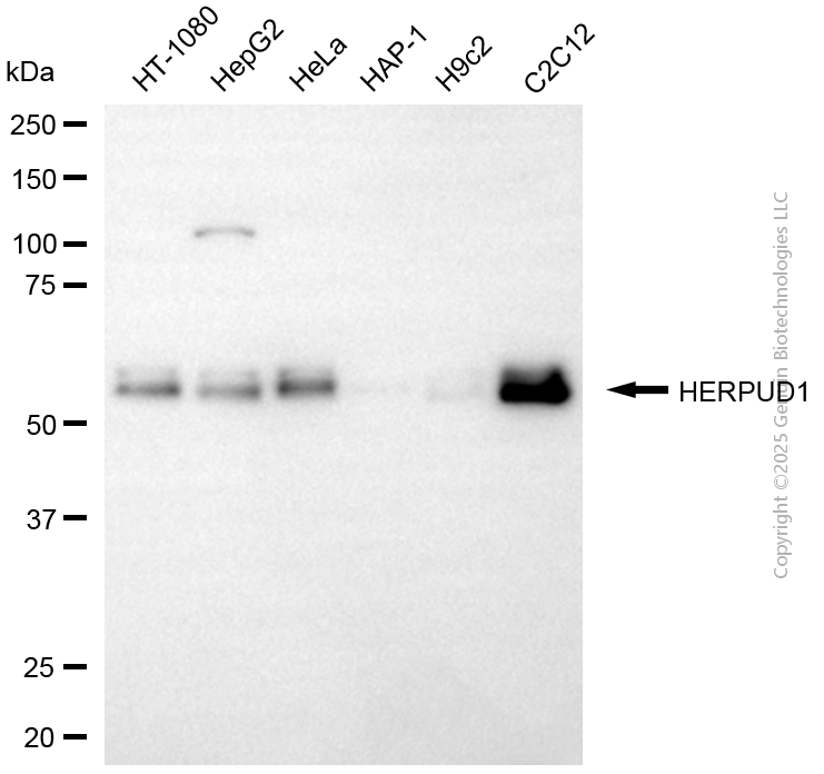
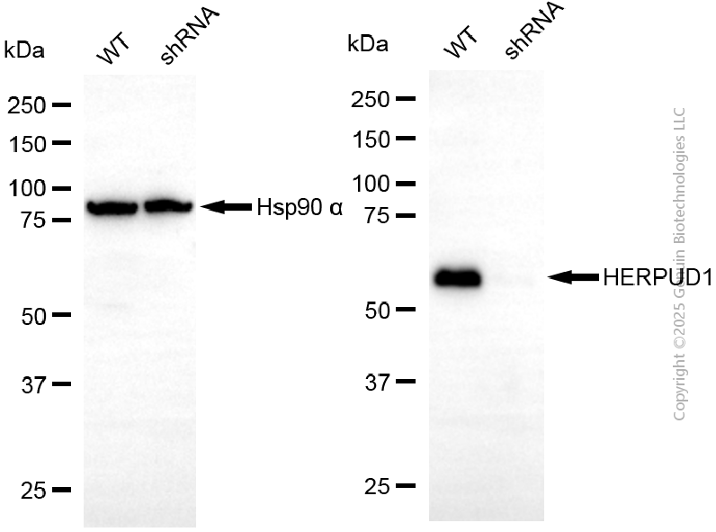
KD-Validated Anti-HERPUD1 Mouse Monoclonal Antibody
Mouse monoclonal Antibody
- SPECIFICATION
- CITATIONS
- PROTOCOLS
- BACKGROUND

Application
| WB |
|---|---|
| Primary Accession | Q15011 |
| Reactivity | Rat, Human, Mouse |
| Clonality | Monoclonal |
| Isotype | Mouse IgG2a |
| Clone Names | 24GB14035 |
| Calculated MW | Predicted, 44 kDa, observed, 54 kDa |
| Gene Name | HERPUD1 |
| Aliases | HERPUD1; Homocysteine Inducible ER Protein With Ubiquitin Like Domain 1; HERP; KIAA0025; Mif1; SUP; Homocysteine-Inducible, Endoplasmic Reticulum Stress-Inducible, Ubiquitin-Like Domain Member 1; Homocysteine-Responsive Endoplasmic Reticulum-Resident Ubiquitin-Like Domain Member 1 Protein; Methyl Methanesulfonate (MMF)-Inducible Fragment Protein 1; omocysteine-Inducible Endoplasmic Reticulum Stress-Inducible Ubiquitin-Like Domain Member 1 Protein; MMS-Inducible; MIF1 |
| Immunogen | Recombinant protein of human HERPUD1 |
| Gene ID | 9709 |
|---|---|
| Other Names | Homocysteine-responsive endoplasmic reticulum-resident ubiquitin-like domain member 1 protein, Methyl methanesulfonate (MMF)-inducible fragment protein 1, HERPUD1, HERP, KIAA0025, MIF1 |
| Name | HERPUD1 |
|---|---|
| Synonyms | HERP, KIAA0025, MIF1 |
| Function | Component of the endoplasmic reticulum quality control (ERQC) system also called ER-associated degradation (ERAD) involved in ubiquitin-dependent degradation of misfolded endoplasmic reticulum proteins (PubMed:16289116, PubMed:28827405). Could enhance presenilin- mediated amyloid-beta protein 40 generation. Binds to ubiquilins and this interaction is required for efficient degradation of CD3D via the ERAD pathway (PubMed:18307982). |
| Cellular Location | Endoplasmic reticulum membrane; Multi-pass membrane protein |
| Tissue Location | Widely expressed; in the brain, expression seems to be restricted to neurons and vascular smooth muscle cells. Present in activated microglia in senile plaques in the brain of patients with Alzheimer disease |
Research Areas
Citations (0)

Thousands of laboratories across the world have published research that depended on the performance of antibodies from Abcepta to advance their research. Check out links to articles that cite our products in major peer-reviewed journals, organized by research category.
Submit your citation using an Abcepta antibody to
info@abcepta.com, and receive a free "I Love Antibodies" mug.
info@abcepta.com, and receive a free "I Love Antibodies" mug.

Application Protocols
Provided below are standard protocols that you may find useful for product applications.
Abcepta welcomes feedback from its customers.
If you have used an Abcepta product and would like to share how it has performed, please click on the "Submit Review" button and provide the requested information. Our staff will examine and post your review and contact you if needed.
If you have any additional inquiries please email technical services at tech@abcepta.com.
$ 399.20

$ 149.00
Cat# AGI2169
Ordering Information
United States
AlbaniaAustraliaAustriaBelgiumBosnia & HerzegovinaBrazilBulgariaCanadaCentral AmericaChinaCroatiaCyprusCzech RepublicDenmarkEstoniaFinlandFranceGermanyGreeceHong KongHungaryIcelandIndiaIndonesiaIrelandIsraelItalyJapanLatviaLithuaniaLuxembourgMacedoniaMalaysiaMaltaMexicoNetherlandsNew ZealandNorwayPakistanPolandPortugalRomaniaSerbiaSingaporeSlovakiaSloveniaSouth AfricaSouth KoreaSpainSwedenSwitzerlandTaiwanTurkeyUnited KingdomUnited StatesVietnamWorldwideOthers
USA Headquarters
(888) 735-7227 / (858) 622-0099 or (858) 875-1900
Other Products
Shipping Information
Domestic orders (in stock items)
Shipped out the same day. Orders placed after 1 PM (PST) will ship out the next business day.
International orders
Contact your local distributors
