


 Foundational characteristics of cancer include proliferation, angiogenesis, migration, evasion of apoptosis, and cellular immortality. Find key markers for these cellular processes and antibodies to detect them.
 The SUMOplot™ Analysis Program predicts and scores sumoylation sites in your protein. SUMOylation is a post-translational modification involved in various cellular processes, such as nuclear-cytosolic transport, transcriptional regulation, apoptosis, protein stability, response to stress, and progression through the cell cycle.
 The Autophagy Receptor Motif Plotter predicts and scores autophagy receptor binding sites in your protein. Identifying proteins connected to this pathway is critical to understanding the role of autophagy in physiological as well as pathological processes such as development, differentiation, neurodegenerative diseases, stress, infection, and cancer.
> home > Products > Primary Antibodies > Antibody Collections > KD-Validated Antibodies > KD-Validated Anti-MYH9 Mouse Monoclonal Antibody
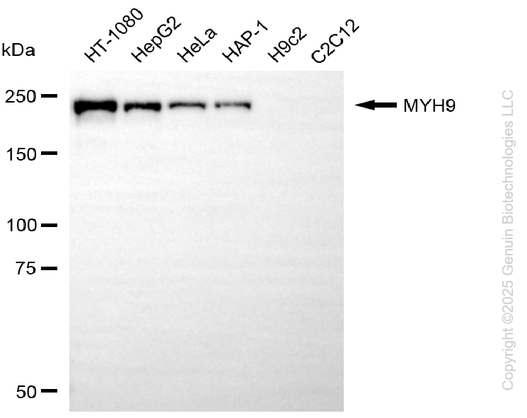
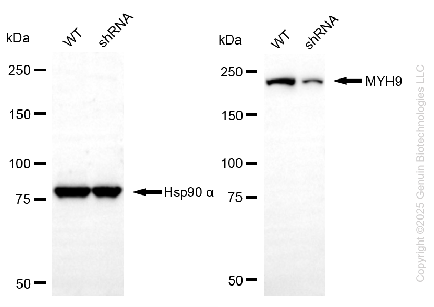
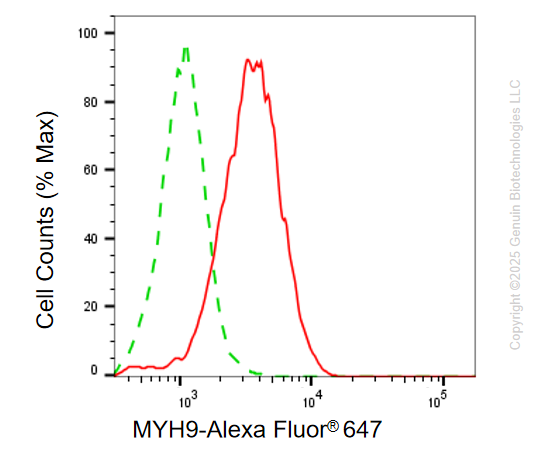
KD-Validated Anti-MYH9 Mouse Monoclonal Antibody
Mouse monoclonal antibody
- SPECIFICATION
- CITATIONS
- PROTOCOLS
- BACKGROUND

Application
| WB, FC |
|---|---|
| Primary Accession | P35579 |
| Reactivity | Human |
| Clonality | Monoclonal |
| Isotype | Mouse IgG2b |
| Clone Names | 24GB14225 |
| Calculated MW | Predicted, 227 kDa, observed, 227 kDa |
| Gene Name | MYH9 |
| Aliases | MYH9; Myosin Heavy Chain 9; NMHC-II-A; NMMHCA; EPSTS; FTNS; MHA; Cellular Myosin Heavy Chain, Type A; Nonmuscle Myosin Heavy Chain II-A; Non-Muscle Myosin Heavy Chain IIa; Non-Muscle Myosin Heavy Chain A; MMHC-IIA; Myosin-9; DFNA17; Myosin, Heavy Polypeptide 9, Non-Muscle; Non-Muscle Myosin Heavy Polypeptide 9; Myosin Heavy Chain, Non-Muscle IIa; Myosin, Heavy Chain 9, Non-Muscle; Non-Muscle Myosin Heavy Chain 9; Nonmuscle Myosin IIA2; NMMHC II-A; NMMHC-A; BDPLT6; MATINS |
| Immunogen | Recombinant protein of human MYH9 |
| Gene ID | 4627 |
|---|---|
| Other Names | Myosin-9, Cellular myosin heavy chain, type A, Myosin heavy chain 9, Myosin heavy chain, non-muscle IIa, Non-muscle myosin heavy chain A, NMMHC-A, Non-muscle myosin heavy chain IIa, NMMHC II-a, NMMHC-IIA, MYH9 |
| Name | MYH9 |
|---|---|
| Function | Cellular myosin that appears to play a role in cytokinesis, cell shape, and specialized functions such as secretion and capping. Required for cortical actin clearance prior to oocyte exocytosis (By similarity). Promotes cell motility in conjunction with S100A4 (PubMed:16707441). During cell spreading, plays an important role in cytoskeleton reorganization, focal contact formation (in the margins but not the central part of spreading cells), and lamellipodial retraction; this function is mechanically antagonized by MYH10 (PubMed:20052411). |
| Cellular Location | Cytoplasm, cytoskeleton. Cytoplasm, cell cortex {ECO:0000250|UniProtKB:Q8VDD5}. Cytoplasmic vesicle, secretory vesicle, Cortical granule {ECO:0000250|UniProtKB:Q8VDD5}. Cell membrane Note=Colocalizes with actin filaments at lamellipodia margins and at the leading edge of migrating cells (PubMed:20052411). In retinal pigment epithelial cells, predominantly localized to stress fiber-like structures with some localization to cytoplasmic puncta (PubMed:27331610). |
| Tissue Location | In the kidney, expressed in the glomeruli. Also expressed in leukocytes. |
Research Areas
Citations (0)

Thousands of laboratories across the world have published research that depended on the performance of antibodies from Abcepta to advance their research. Check out links to articles that cite our products in major peer-reviewed journals, organized by research category.
Submit your citation using an Abcepta antibody to
info@abcepta.com, and receive a free "I Love Antibodies" mug.
info@abcepta.com, and receive a free "I Love Antibodies" mug.

Application Protocols
Provided below are standard protocols that you may find useful for product applications.
Abcepta welcomes feedback from its customers.
If you have used an Abcepta product and would like to share how it has performed, please click on the "Submit Review" button and provide the requested information. Our staff will examine and post your review and contact you if needed.
If you have any additional inquiries please email technical services at tech@abcepta.com.
$ 399.20

$ 149.00
Cat# AGI2177
Ordering Information
United States
AlbaniaAustraliaAustriaBelgiumBosnia & HerzegovinaBrazilBulgariaCanadaCentral AmericaChinaCroatiaCyprusCzech RepublicDenmarkEstoniaFinlandFranceGermanyGreeceHong KongHungaryIcelandIndiaIndonesiaIrelandIsraelItalyJapanLatviaLithuaniaLuxembourgMacedoniaMalaysiaMaltaMexicoNetherlandsNew ZealandNorwayPakistanPolandPortugalRomaniaSerbiaSingaporeSlovakiaSloveniaSouth AfricaSouth KoreaSpainSwedenSwitzerlandTaiwanTurkeyUnited KingdomUnited StatesVietnamWorldwideOthers
USA Headquarters
(888) 735-7227 / (858) 622-0099 or (858) 875-1900
Other Products
Shipping Information
Domestic orders (in stock items)
Shipped out the same day. Orders placed after 1 PM (PST) will ship out the next business day.
International orders
Contact your local distributors
