


 Foundational characteristics of cancer include proliferation, angiogenesis, migration, evasion of apoptosis, and cellular immortality. Find key markers for these cellular processes and antibodies to detect them.
 The SUMOplot™ Analysis Program predicts and scores sumoylation sites in your protein. SUMOylation is a post-translational modification involved in various cellular processes, such as nuclear-cytosolic transport, transcriptional regulation, apoptosis, protein stability, response to stress, and progression through the cell cycle.
 The Autophagy Receptor Motif Plotter predicts and scores autophagy receptor binding sites in your protein. Identifying proteins connected to this pathway is critical to understanding the role of autophagy in physiological as well as pathological processes such as development, differentiation, neurodegenerative diseases, stress, infection, and cancer.
> home > Products > Primary Antibodies > Antibody Collections > KD-Validated Antibodies > KD-Validated Anti-TRIM34 Mouse Monoclonal Antibody
KD-Validated Anti-TRIM34 Mouse Monoclonal Antibody
Mouse monoclonal antibody
- SPECIFICATION
- CITATIONS
- PROTOCOLS
- BACKGROUND

Application
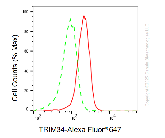
| WB, FC |
|---|---|
| Primary Accession | Q9BYJ4 |
| Reactivity | Human |
| Clonality | Monoclonal |
| Isotype | Mouse IgG2a |
| Clone Names | 24GB14230 |
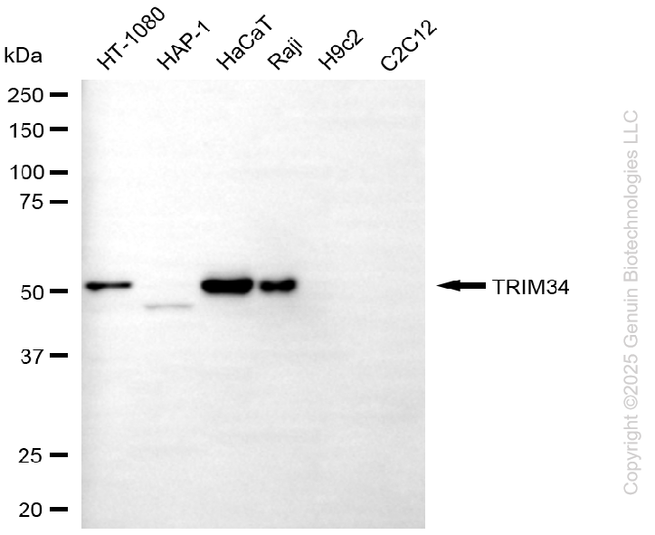
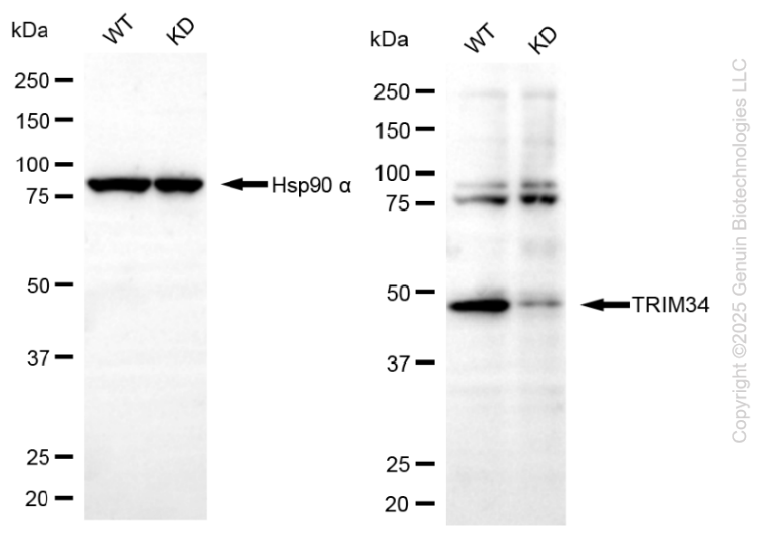
| Calculated MW | Predicted, 57 kDa, observed, 51 kDa |
| Gene Name | TRIM34 |
| Aliases | TRIM34; Tripartite Motif Containing 34; RNF21; Interferon-Responsive Finger Protein 1; E3 Ubiquitin-Protein Ligase TRIM34; IFP1; Ring Finger Protein 21, Interferon-Responsive; Tripartite Motif-Containing 34; RING Finger Protein 21; EC 2.3.2.27; EC 6.3.2 |
| Immunogen | Recombinant protein of human TRIM34 |
| Gene ID | 53840 |
|---|---|
| Other Names | E3 ubiquitin-protein ligase TRIM34, 2.3.2.27, Interferon-responsive finger protein 1, RING finger protein 21, TRIM34, IFP1, RNF21 |
| Name | TRIM34 |
|---|---|
| Synonyms | IFP1, RNF21 |
| Function | Functions as antiviral protein and contributes to the defense against retroviral infections (PubMed:17156811, PubMed:32282853). Acts as a capsid-specific restriction factor with the help of TRIM5 and prevents infection from non-host-adapted retroviruses (PubMed:32282853). During influenza A virus infection, promotes programmed cell death by targeting ZBP1 for 'Lys-63'-linked polyubiquitination (PubMed:35065966). In turn, promotes ZBP1 recruitment of RIPK3 to mediate virus-induced programmed necrosis (PubMed:35065966). Negatively regulates the function of mitochondria by enhancing mitochondrial depolarization leading to cytochrome c release and mitochondria-dependent apoptosis (PubMed:31956709). Also promotes the formation of multinucleated giant cells by means of cell fusion and phagocytosis in epithelial cells (PubMed:31487507). |
| Cellular Location | Cytoplasm Mitochondrion. Note=Localizes in cytoplasmic bodies together with TRIM5 and incoming HIV-1 capsids during infection. |
| Tissue Location | [Isoform 1]: Is the most abundant form. It is highly expressed in the placenta, spleen, colon and peripheral blood leukocytes. |
Research Areas
Citations (0)

Thousands of laboratories across the world have published research that depended on the performance of antibodies from Abcepta to advance their research. Check out links to articles that cite our products in major peer-reviewed journals, organized by research category.
Submit your citation using an Abcepta antibody to
info@abcepta.com, and receive a free "I Love Antibodies" mug.
info@abcepta.com, and receive a free "I Love Antibodies" mug.

Application Protocols
Provided below are standard protocols that you may find useful for product applications.
Abcepta welcomes feedback from its customers.
If you have used an Abcepta product and would like to share how it has performed, please click on the "Submit Review" button and provide the requested information. Our staff will examine and post your review and contact you if needed.
If you have any additional inquiries please email technical services at tech@abcepta.com.
$ 399.20

$ 149.00
Cat# AGI2178
Ordering Information
United States
AlbaniaAustraliaAustriaBelgiumBosnia & HerzegovinaBrazilBulgariaCanadaCentral AmericaChinaCroatiaCyprusCzech RepublicDenmarkEstoniaFinlandFranceGermanyGreeceHong KongHungaryIcelandIndiaIndonesiaIrelandIsraelItalyJapanLatviaLithuaniaLuxembourgMacedoniaMalaysiaMaltaMexicoNetherlandsNew ZealandNorwayPakistanPolandPortugalRomaniaSerbiaSingaporeSlovakiaSloveniaSouth AfricaSouth KoreaSpainSwedenSwitzerlandTaiwanTurkeyUnited KingdomUnited StatesVietnamWorldwideOthers
USA Headquarters
(888) 735-7227 / (858) 622-0099 or (858) 875-1900
Other Products
Shipping Information
Domestic orders (in stock items)
Shipped out the same day. Orders placed after 1 PM (PST) will ship out the next business day.
International orders
Contact your local distributors
