


 Foundational characteristics of cancer include proliferation, angiogenesis, migration, evasion of apoptosis, and cellular immortality. Find key markers for these cellular processes and antibodies to detect them.
 The SUMOplot™ Analysis Program predicts and scores sumoylation sites in your protein. SUMOylation is a post-translational modification involved in various cellular processes, such as nuclear-cytosolic transport, transcriptional regulation, apoptosis, protein stability, response to stress, and progression through the cell cycle.
 The Autophagy Receptor Motif Plotter predicts and scores autophagy receptor binding sites in your protein. Identifying proteins connected to this pathway is critical to understanding the role of autophagy in physiological as well as pathological processes such as development, differentiation, neurodegenerative diseases, stress, infection, and cancer.
> home > Products > Primary Antibodies > Antibody Collections > SARS-Human interaction partners > KD-Validated Anti-DAB2 Rabbit Monoclonal Antibody
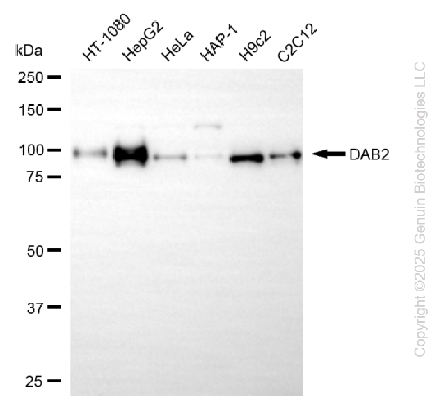
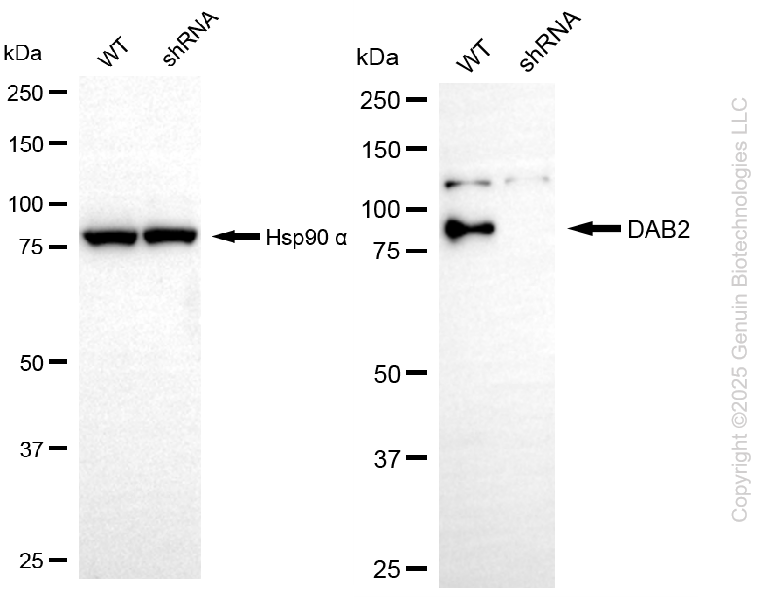
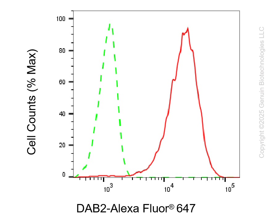
KD-Validated Anti-DAB2 Rabbit Monoclonal Antibody
Rabbit monoclonal Antibody
- SPECIFICATION
- CITATIONS
- PROTOCOLS
- BACKGROUND

Application
| WB, FC |
|---|---|
| Primary Accession | P98082 |
| Reactivity | Rat, Human, Mouse |
| Clonality | Monoclonal |
| Isotype | Rabbit IgG |
| Clone Names | 25GB225 |
| Calculated MW | Predicted, 82 kDa, observed, 96 kDa |
| Gene Name | DAB2 |
| Aliases | DAB2; DAB Adaptor Protein 2; DOC-2; Differentially Expressed In Ovarian Carcinoma 2; Differentially-Expressed Protein 2; DAB2, Clathrin Adaptor Protein 2; Adaptor Molecule Disabled-2; Disabled Homolog 2; DOC2; Disabled (Drosophila) Homolog 2 (Mitogen-Responsive Phosphoprotein); Disabled Homolog 2, Mitogen-Responsive Phosphoprotein (Drosophila); Dab, Mitogen-Responsive Phosphoprotein, Homolog 2 (Drosophila); Disabled Homolog 2, Mitogen-Responsive Phosphoprotein; Dab, Mitogen-Responsive Phosphoprotein, Homolog 2 |
| Immunogen | A synthesized peptide derived from human DAB2 |
| Gene ID | 1601 |
|---|---|
| Other Names | Disabled homolog 2, Adaptor molecule disabled-2, Differentially expressed in ovarian carcinoma 2, DOC-2, Differentially-expressed protein 2, DAB2, DOC2 |
| Name | DAB2 |
|---|---|
| Synonyms | DOC2 |
| Function | Adapter protein that functions as a clathrin-associated sorting protein (CLASP) required for clathrin-mediated endocytosis of selected cargo proteins. Can bind and assemble clathrin, and binds simultaneously to phosphatidylinositol 4,5-bisphosphate (PtdIns(4,5)P2) and cargos containing non-phosphorylated NPXY internalization motifs, such as the LDL receptor, to recruit them to clathrin-coated pits. Can function in clathrin-mediated endocytosis independently of the AP-2 complex. Involved in endocytosis of integrin beta-1; this function seems to redundant with the AP-2 complex and seems to require DAB2 binding to endocytosis accessory EH domain-containing proteins such as EPS15, EPS15L1 and ITSN1. Involved in endocytosis of cystic fibrosis transmembrane conductance regulator/CFTR. Involved in endocytosis of megalin/LRP2 lipoprotein receptor during embryonal development. Required for recycling of the TGF-beta receptor. Involved in CFTR trafficking to the late endosome. Involved in several receptor-mediated signaling pathways. Involved in TGF-beta receptor signaling and facilitates phosphorylation of the signal transducer SMAD2. Mediates TFG-beta-stimulated JNK activation. May inhibit the canoniocal Wnt/beta-catenin signaling pathway by stabilizing the beta-catenin destruction complex through a competing association with axin preventing its dephosphorylation through protein phosphatase 1 (PP1). Sequesters LRP6 towards clathrin-mediated endocytosis, leading to inhibition of Wnt/beta-catenin signaling. May activate non-canonical Wnt signaling. In cell surface growth factor/Ras signaling pathways proposed to inhibit ERK activation by interrupting the binding of GRB2 to SOS1 and to inhibit SRC by preventing its activating phosphorylation at 'Tyr-419'. Proposed to be involved in modulation of androgen receptor (AR) signaling mediated by SRC activation; seems to compete with AR for interaction with SRC. Plays a role in the CSF-1 signal transduction pathway. Plays a role in cellular differentiation. Involved in cell positioning and formation of visceral endoderm (VE) during embryogenesis and proposed to be required in the VE to respond to Nodal signaling coming from the epiblast. Required for the epithelial to mesenchymal transition, a process necessary for proper embryonic development. May be involved in myeloid cell differentiation and can induce macrophage adhesion and spreading. May act as a tumor suppressor. |
| Cellular Location | Cytoplasm. Cytoplasmic vesicle, clathrin-coated vesicle membrane. Membrane, clathrin-coated pit. Note=Colocalizes with large insert-containing isoforms of MYO6 at clathrin-coated pits/vesicles. During mitosis is progressively displaced from the membrane and translocated to the cytoplasm |
| Tissue Location | Expressed in deep invaginations, inclusion cysts and the surface epithelial cells of the ovary. Also expressed in breast epithelial cells, spleen, thymus, prostate, testis, macrophages, fibroblasts, lung epithelial cells, placenta, brain stem, heart and small intestine. Expressed in kidney proximal tubular epithelial cells (at protein level). |
Research Areas
Citations (0)

Thousands of laboratories across the world have published research that depended on the performance of antibodies from Abcepta to advance their research. Check out links to articles that cite our products in major peer-reviewed journals, organized by research category.
Submit your citation using an Abcepta antibody to
info@abcepta.com, and receive a free "I Love Antibodies" mug.
info@abcepta.com, and receive a free "I Love Antibodies" mug.

Application Protocols
Provided below are standard protocols that you may find useful for product applications.
Abcepta welcomes feedback from its customers.
If you have used an Abcepta product and would like to share how it has performed, please click on the "Submit Review" button and provide the requested information. Our staff will examine and post your review and contact you if needed.
If you have any additional inquiries please email technical services at tech@abcepta.com.
$ 399.20

$ 149.00
Cat# AGI2209
Ordering Information
United States
AlbaniaAustraliaAustriaBelgiumBosnia & HerzegovinaBrazilBulgariaCanadaCentral AmericaChinaCroatiaCyprusCzech RepublicDenmarkEstoniaFinlandFranceGermanyGreeceHong KongHungaryIcelandIndiaIndonesiaIrelandIsraelItalyJapanLatviaLithuaniaLuxembourgMacedoniaMalaysiaMaltaMexicoNetherlandsNew ZealandNorwayPakistanPolandPortugalRomaniaSerbiaSingaporeSlovakiaSloveniaSouth AfricaSouth KoreaSpainSwedenSwitzerlandTaiwanTurkeyUnited KingdomUnited StatesVietnamWorldwideOthers
USA Headquarters
(888) 735-7227 / (858) 622-0099 or (858) 875-1900
Other Products
Shipping Information
Domestic orders (in stock items)
Shipped out the same day. Orders placed after 1 PM (PST) will ship out the next business day.
International orders
Contact your local distributors
