


 Foundational characteristics of cancer include proliferation, angiogenesis, migration, evasion of apoptosis, and cellular immortality. Find key markers for these cellular processes and antibodies to detect them.
 The SUMOplot™ Analysis Program predicts and scores sumoylation sites in your protein. SUMOylation is a post-translational modification involved in various cellular processes, such as nuclear-cytosolic transport, transcriptional regulation, apoptosis, protein stability, response to stress, and progression through the cell cycle.
 The Autophagy Receptor Motif Plotter predicts and scores autophagy receptor binding sites in your protein. Identifying proteins connected to this pathway is critical to understanding the role of autophagy in physiological as well as pathological processes such as development, differentiation, neurodegenerative diseases, stress, infection, and cancer.
> home > Products > Primary Antibodies > Antibody Collections > KD-Validated Antibodies > KD-Validated Anti-Fibronectin 1 Rabbit Monoclonal Antibody
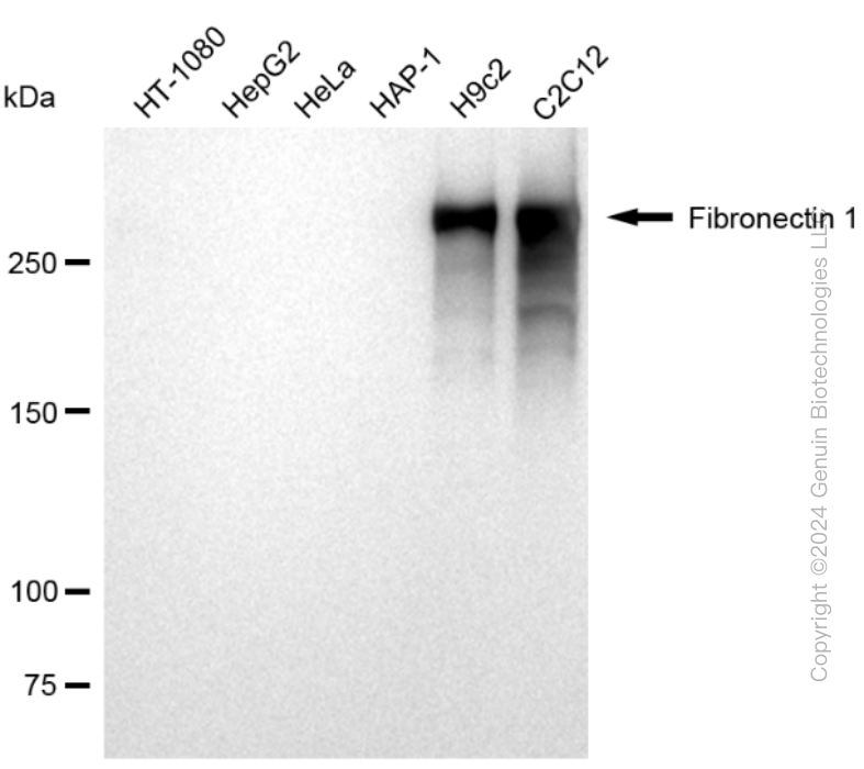
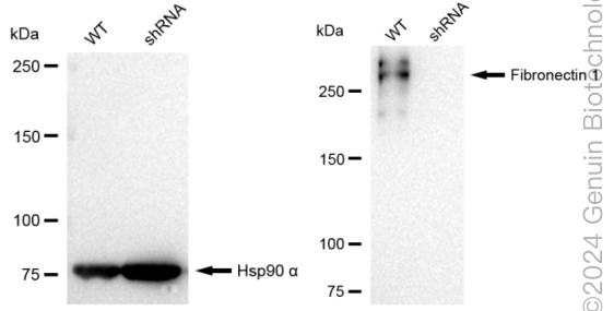
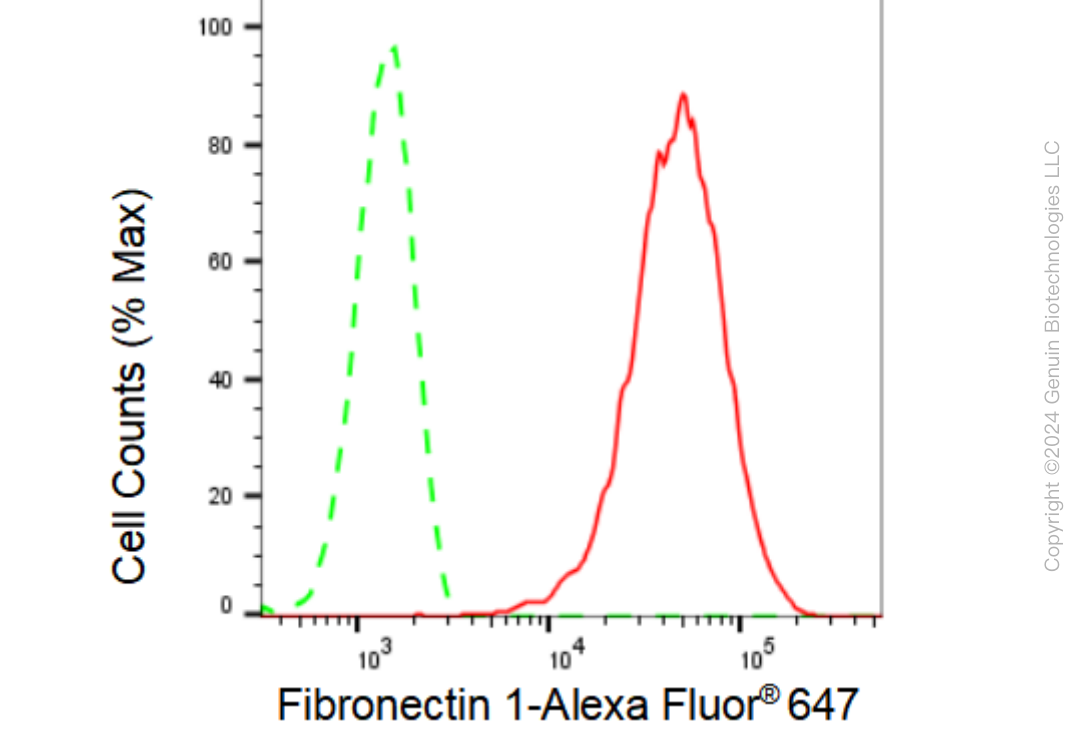
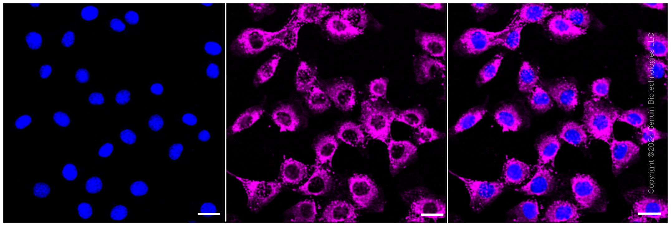
KD-Validated Anti-Fibronectin 1 Rabbit Monoclonal Antibody
Rabbit monoclonal antibody
- SPECIFICATION
- CITATIONS
- PROTOCOLS
- BACKGROUND

Application
| WB, FC, ICC |
|---|---|
| Primary Accession | P02751 |
| Reactivity | Rat, Human, Mouse |
| Clonality | Monoclonal |
| Isotype | Rabbit IgG |
| Clone Names | 23GB1270 |
| Calculated MW | Predicted, 263 kDa ; Observed, 263 kDa |
| Gene Name | FN1 |
| Aliases | Fibronectin1; CIG; Cold-Insoluble Globulin; GFND2; LETS ; FINC; MSF; Migration-Stimulating Factor; Lnc-ABCA12-8; Fibronectin; FN; Epididymis Secretory Sperm Binding Protein; LNC-ABCA12-8; SMDCF; ED-B; GFND; FNZ |
| Immunogen | A synthesized peptide derived from human Fibronectin |
| Gene ID | 2335 |
|---|---|
| Other Names | Fibronectin, FN, Cold-insoluble globulin, CIG, Anastellin, Ugl-Y1, Ugl-Y2, Ugl-Y3, FN1 (HGNC:3778), FN |
| Name | FN1 (HGNC:3778) |
|---|---|
| Synonyms | FN |
| Function | Fibronectins bind cell surfaces and various compounds including collagen, fibrin, heparin, DNA, and actin (PubMed:3024962, PubMed:3593230, PubMed:3900070, PubMed:7989369). Fibronectins are involved in cell adhesion, cell motility, opsonization, wound healing, and maintenance of cell shape (PubMed:3024962, PubMed:3593230, PubMed:3900070, PubMed:7989369). Involved in osteoblast compaction through the fibronectin fibrillogenesis cell-mediated matrix assembly process, essential for osteoblast mineralization (By similarity). Participates in the regulation of type I collagen deposition by osteoblasts (By similarity). Acts as a ligand for the LILRB4 receptor, inhibiting FCGR1A/CD64-mediated monocyte activation (PubMed:34089617). |
| Cellular Location | Secreted, extracellular space, extracellular matrix. Secreted {ECO:0000250|UniProtKB:P11276} |
| Tissue Location | Expressed in the inner limiting membrane and around blood vessels in the retina (at protein level) (PubMed:29777959) Plasma FN (soluble dimeric form) is secreted by hepatocytes. Cellular FN (dimeric or cross-linked multimeric forms), made by fibroblasts, epithelial and other cell types, is deposited as fibrils in the extracellular matrix. Ugl-Y1, Ugl-Y2 and Ugl-Y3 are found in urine (PubMed:17614963). |
Research Areas
Citations (0)

Thousands of laboratories across the world have published research that depended on the performance of antibodies from Abcepta to advance their research. Check out links to articles that cite our products in major peer-reviewed journals, organized by research category.
Submit your citation using an Abcepta antibody to
info@abcepta.com, and receive a free "I Love Antibodies" mug.
info@abcepta.com, and receive a free "I Love Antibodies" mug.

Application Protocols
Provided below are standard protocols that you may find useful for product applications.
Abcepta welcomes feedback from its customers.
If you have used an Abcepta product and would like to share how it has performed, please click on the "Submit Review" button and provide the requested information. Our staff will examine and post your review and contact you if needed.
If you have any additional inquiries please email technical services at tech@abcepta.com.
$ 399.20

$ 149.00
Cat# AGI2367
Ordering Information
United States
AlbaniaAustraliaAustriaBelgiumBosnia & HerzegovinaBrazilBulgariaCanadaCentral AmericaChinaCroatiaCyprusCzech RepublicDenmarkEstoniaFinlandFranceGermanyGreeceHong KongHungaryIcelandIndiaIndonesiaIrelandIsraelItalyJapanLatviaLithuaniaLuxembourgMacedoniaMalaysiaMaltaMexicoNetherlandsNew ZealandNorwayPakistanPolandPortugalRomaniaSerbiaSingaporeSlovakiaSloveniaSouth AfricaSouth KoreaSpainSwedenSwitzerlandTaiwanTurkeyUnited KingdomUnited StatesVietnamWorldwideOthers
USA Headquarters
(888) 735-7227 / (858) 622-0099 or (858) 875-1900
Other Products
Shipping Information
Domestic orders (in stock items)
Shipped out the same day. Orders placed after 1 PM (PST) will ship out the next business day.
International orders
Contact your local distributors
