


 Foundational characteristics of cancer include proliferation, angiogenesis, migration, evasion of apoptosis, and cellular immortality. Find key markers for these cellular processes and antibodies to detect them.
 The SUMOplot™ Analysis Program predicts and scores sumoylation sites in your protein. SUMOylation is a post-translational modification involved in various cellular processes, such as nuclear-cytosolic transport, transcriptional regulation, apoptosis, protein stability, response to stress, and progression through the cell cycle.
 The Autophagy Receptor Motif Plotter predicts and scores autophagy receptor binding sites in your protein. Identifying proteins connected to this pathway is critical to understanding the role of autophagy in physiological as well as pathological processes such as development, differentiation, neurodegenerative diseases, stress, infection, and cancer.
MART-1 / Melan-A / MLANA (Melanoma Marker) Antibody - With BSA and Azide
Mouse Monoclonal Antibody [Clone SPM342 ]
- SPECIFICATION
- CITATIONS
- PROTOCOLS
- BACKGROUND

Application
| WB, IHC-P, IF, FC |
|---|---|
| Primary Accession | Q16655 |
| Other Accession | 2315, 154069 |
| Reactivity | Human, Horse |
| Host | Mouse |
| Clonality | Monoclonal |
| Isotype | Mouse / IgG2b, kappa |
| Clone Names | SPM342 |
| Calculated MW | 20-22kDa (doublet) |
| Gene ID | 2315 |
|---|---|
| Other Names | Melanoma antigen recognized by T-cells 1, MART-1, Antigen LB39-AA, Antigen SK29-AA, Protein Melan-A, MLANA, MART1 |
| Application Note | WB~~1:1000 IHC-P~~N/A IF~~1:50~200 FC~~1:10~50 |
| Format | 200ug/ml of Ab purified from Bioreactor Concentrate by Protein A/G. Prepared in 10mM PBS with 0.05% BSA & 0.05% azide. Also available WITHOUT BSA & azide at 1.0mg/ml. |
| Storage | Store at 2 to 8°C.Antibody is stable for 24 months. |
| Precautions | MART-1 / Melan-A / MLANA (Melanoma Marker) Antibody - With BSA and Azide is for research use only and not for use in diagnostic or therapeutic procedures. |
| Name | MLANA |
|---|---|
| Synonyms | MART1 |
| Function | Involved in melanosome biogenesis by ensuring the stability of GPR143. Plays a vital role in the expression, stability, trafficking, and processing of melanocyte protein PMEL, which is critical to the formation of stage II melanosomes. |
| Cellular Location | Endoplasmic reticulum membrane; Single-pass type III membrane protein. Golgi apparatus. Golgi apparatus, trans-Golgi network membrane. Melanosome. Note=Also found in small vesicles and tubules dispersed over the entire cytoplasm. A small fraction of the protein is inserted into the membrane in an inverted orientation Inversion of membrane topology results in the relocalization of the protein from a predominant Golgi/post-Golgi area to the endoplasmic reticulum. Melanoma cells expressing the protein with an inverted membrane topology are more effectively recognized by specific cytolytic T-lymphocytes than those expressing the protein in its native membrane orientation |
| Tissue Location | Expression is restricted to melanoma and melanocyte cell lines and retina |

Thousands of laboratories across the world have published research that depended on the performance of antibodies from Abcepta to advance their research. Check out links to articles that cite our products in major peer-reviewed journals, organized by research category.
info@abcepta.com, and receive a free "I Love Antibodies" mug.

Provided below are standard protocols that you may find useful for product applications.
Background
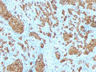
This antibody recognizes a protein doublet of 20-22kDa, identified as MART-1 (Melanoma Antigen Recognized by T cells 1) or Melan-A. MART-1 is a newly identified melanocyte differentiation antigen recognized by autologous cytotoxic T lymphocytes. Seven other melanoma associated antigens recognized by autologous cytotoxic T cells include MAGE-1, MAGE-3, tyrosinase, gp100, gp75, BAGE-1, and GAGE-1. Subcellular fractionation shows that MART-1 is present in melanosomes and endoplasmic reticulum. This MAb labels melanomas and other tumors showing melanocytic differentiation. It is also a useful positive-marker for angiomyolipomas. It does not stain tumor cells of epithelial, lymphoid, glial, or mesenchymal origin.
References
Kawakami Y, et. al. Journal of Immunological Methods, 1997, 202(1):13-25. | Marincola FM, et. al. Journal of Immunotherapy with Emphasis on Tumor Immunology, 1996, 19(3):192-205
If you have used an Abcepta product and would like to share how it has performed, please click on the "Submit Review" button and provide the requested information. Our staff will examine and post your review and contact you if needed.
If you have any additional inquiries please email technical services at tech@abcepta.com.
Ordering Information
Other Products
Shipping Information
