


 Foundational characteristics of cancer include proliferation, angiogenesis, migration, evasion of apoptosis, and cellular immortality. Find key markers for these cellular processes and antibodies to detect them.
 The SUMOplot™ Analysis Program predicts and scores sumoylation sites in your protein. SUMOylation is a post-translational modification involved in various cellular processes, such as nuclear-cytosolic transport, transcriptional regulation, apoptosis, protein stability, response to stress, and progression through the cell cycle.
 The Autophagy Receptor Motif Plotter predicts and scores autophagy receptor binding sites in your protein. Identifying proteins connected to this pathway is critical to understanding the role of autophagy in physiological as well as pathological processes such as development, differentiation, neurodegenerative diseases, stress, infection, and cancer.
CD50 / ICAM-3 Antibody - With BSA and Azide
Mouse Monoclonal Antibody [Clone SPM505 ]
- SPECIFICATION
- CITATIONS
- PROTOCOLS
- BACKGROUND

Application
| IHC-P, IF, FC |
|---|---|
| Primary Accession | P32942 |
| Other Accession | 3385, 354563 |
| Reactivity | Human |
| Host | Mouse |
| Clonality | Monoclonal |
| Isotype | Mouse / IgG2b, kappa |
| Clone Names | SPM505 |
| Calculated MW | 110-160kDa |
| Gene ID | 3385 |
|---|---|
| Other Names | Intercellular adhesion molecule 3, ICAM-3, CDw50, ICAM-R, CD50, ICAM3 |
| Application Note | IHC-P~~N/A IF~~1:50~200 FC~~1:10~50 |
| Format | 200ug/ml of Ab purified from Bioreactor Concentrate by Protein A/G. Prepared in 10mM PBS with 0.05% BSA & 0.05% azide. Also available WITHOUT BSA & azide at 1.0mg/ml. |
| Storage | Store at 2 to 8°C.Antibody is stable for 24 months. |
| Precautions | CD50 / ICAM-3 Antibody - With BSA and Azide is for research use only and not for use in diagnostic or therapeutic procedures. |
| Name | ICAM3 |
|---|---|
| Function | ICAM proteins are ligands for the leukocyte adhesion protein LFA-1 (integrin alpha-L/beta-2) (PubMed:1448173). ICAM3 is also a ligand for integrin alpha-D/beta-2. In association with integrin alpha- L/beta-2, contributes to apoptotic neutrophil phagocytosis by macrophages (PubMed:23775590). |
| Cellular Location | Membrane; Single-pass type I membrane protein. |
| Tissue Location | Leukocytes. |

Thousands of laboratories across the world have published research that depended on the performance of antibodies from Abcepta to advance their research. Check out links to articles that cite our products in major peer-reviewed journals, organized by research category.
info@abcepta.com, and receive a free "I Love Antibodies" mug.

Provided below are standard protocols that you may find useful for product applications.
Background
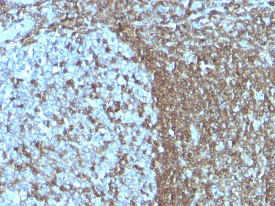
Recognizes the D1 domain of an N-glycosylated glycoprotein of 120kDa with intra-chain disulfide bonds, identified as CD50 or ICAM-3. CD50 is the major ligand for LFA-1 (CD11a/CD18) and may have signaling role to increase adhesion. It is expressed on thymocytes and T lymphocytes and is resistant to treatment with phosphatidylinositol (PI) phospholipase C. This MAb is excellent for staining of formalin/paraffin tissues.
References
LeukocyteTyping V (S.F. Schlossman, et al, eds.) Oxford University Press, Oxford (1995)p. 1546-1547, 1578-1579. | C.L. Holness, et al, (1995) J Biol Chem 270: 877-884. | Y. van Kooyk,et al, (1996) J Exp Med 183:1247-1252. | Leukocyte TypingVI (T. Kishimoto, et al, eds.) GarlandPublishing, Inc., New York (1997) p.403-409
If you have used an Abcepta product and would like to share how it has performed, please click on the "Submit Review" button and provide the requested information. Our staff will examine and post your review and contact you if needed.
If you have any additional inquiries please email technical services at tech@abcepta.com.
Ordering Information
Other Products
Shipping Information
