


 Foundational characteristics of cancer include proliferation, angiogenesis, migration, evasion of apoptosis, and cellular immortality. Find key markers for these cellular processes and antibodies to detect them.
 The SUMOplot™ Analysis Program predicts and scores sumoylation sites in your protein. SUMOylation is a post-translational modification involved in various cellular processes, such as nuclear-cytosolic transport, transcriptional regulation, apoptosis, protein stability, response to stress, and progression through the cell cycle.
 The Autophagy Receptor Motif Plotter predicts and scores autophagy receptor binding sites in your protein. Identifying proteins connected to this pathway is critical to understanding the role of autophagy in physiological as well as pathological processes such as development, differentiation, neurodegenerative diseases, stress, infection, and cancer.
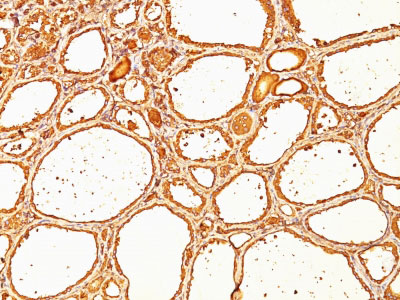
Thyroglobulin (Thyroidal Cell Marker) Antibody - With BSA and Azide
Mouse Monoclonal Antibody [Clone SPM517 ]
- SPECIFICATION
- CITATIONS
- PROTOCOLS
- BACKGROUND

Application
| IHC-P, FC |
|---|---|
| Primary Accession | P01266 |
| Other Accession | 7038, 654591 |
| Reactivity | Human, Mouse, Rat |
| Host | Mouse |
| Clonality | Monoclonal |
| Isotype | Mouse / IgG1, kappa |
| Clone Names | SPM517 |
| Calculated MW | 660kDa (Dimeric Form) |
| Gene ID | 7038 |
|---|---|
| Other Names | Thyroglobulin, Tg, TG |
| Application Note | IHC-P~~N/A FC~~1:10~50 |
| Format | 200ug/ml of Ab purified from Bioreactor Concentrate by Protein A/G. Prepared in 10mM PBS with 0.05% BSA & 0.05% azide. Also available WITHOUT BSA & azide at 1.0mg/ml. |
| Storage | Store at 2 to 8°C.Antibody is stable for 24 months. |
| Precautions | Thyroglobulin (Thyroidal Cell Marker) Antibody - With BSA and Azide is for research use only and not for use in diagnostic or therapeutic procedures. |
| Name | TG (HGNC:11764) |
|---|---|
| Function | Acts as a substrate for the production of iodinated thyroid hormones thyroxine (T4) and triiodothyronine (T3) (PubMed:17532758, PubMed:32025030). The synthesis of T3 and T4 involves iodination of selected tyrosine residues of TG/thyroglobulin followed by their oxidative coupling in the thyroid follicle lumen (PubMed:32025030). Following TG re-internalization and lysosomal-mediated proteolysis, T3 and T4 are released from the polypeptide backbone leading to their secretion into the bloodstream (PubMed:32025030). One dimer produces 7 thyroid hormone molecules (PubMed:32025030). |
| Cellular Location | Secreted. Note=Secreted into the thyroid follicle lumen (PubMed:19509106). Localizes to colloid globules, a structure formed in the thyroid follicle lumen consisting of cross-linked TG arranged in concentric layers (PubMed:11082042, PubMed:8626858). |
| Tissue Location | Specifically expressed in the thyroid gland. |

Thousands of laboratories across the world have published research that depended on the performance of antibodies from Abcepta to advance their research. Check out links to articles that cite our products in major peer-reviewed journals, organized by research category.
info@abcepta.com, and receive a free "I Love Antibodies" mug.

Provided below are standard protocols that you may find useful for product applications.
Background
Thyroglobulin is a 660kDa dimeric pre-protein with mutiple glycosylation sites. It is produced by and processed within the thyroid gland to produce the hormone thyroxine and triiodothyronine. ĀPrior to forming dimers, thyroglobulin monomers undergo conformational maturation in the endoplasmic reticulation. ĀThe vast majority of follicular carcinomas of the thyroid will give positive immunoreactivity for anti-thyroglobulin even though sometimes only focally. Poorly differentiated carcinomas of the thyroid are frequently anti-thyroglobulin negative. Adenocarcinomas of other-than-thyroid origin do not react with this antibody. This antibody is useful in identification of thyroid carcinoma of the papillary and follicular types. Presence of thyroglobulin in metastatic lesions establishes the thyroid origin of tumor. Anti-thyroglobulin, combined with anti-calcitonin, can identify medullary carcinomas of the thyroid. Furthermore, anti-thyroglobulin, combined with anti-TTF1, can be a reliable marker to differentiate between primary thyroid and lung neoplasms.
References
Ossendorp FA, et. al. Journal of Immunological Methods, 1989, 120(2):191-200. | Bellet, D, et al. J Clin Endocrin Metab 1983;56:530-533 | Heffess CS et al. Cancer. 2002;95(9):1869-78 | Judkins AR et al. Hum Pathol. 1999;30(11):1373-
If you have used an Abcepta product and would like to share how it has performed, please click on the "Submit Review" button and provide the requested information. Our staff will examine and post your review and contact you if needed.
If you have any additional inquiries please email technical services at tech@abcepta.com.
Ordering Information
Other Products
Shipping Information
