


 Foundational characteristics of cancer include proliferation, angiogenesis, migration, evasion of apoptosis, and cellular immortality. Find key markers for these cellular processes and antibodies to detect them.
 The SUMOplot™ Analysis Program predicts and scores sumoylation sites in your protein. SUMOylation is a post-translational modification involved in various cellular processes, such as nuclear-cytosolic transport, transcriptional regulation, apoptosis, protein stability, response to stress, and progression through the cell cycle.
 The Autophagy Receptor Motif Plotter predicts and scores autophagy receptor binding sites in your protein. Identifying proteins connected to this pathway is critical to understanding the role of autophagy in physiological as well as pathological processes such as development, differentiation, neurodegenerative diseases, stress, infection, and cancer.
TOX3 / TNRC9 Antibody - With BSA and Azide
Mouse Monoclonal Antibody [Clone TOX3/1123 ]
- SPECIFICATION
- CITATIONS
- PROTOCOLS
- BACKGROUND

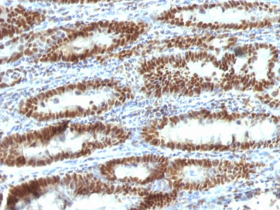
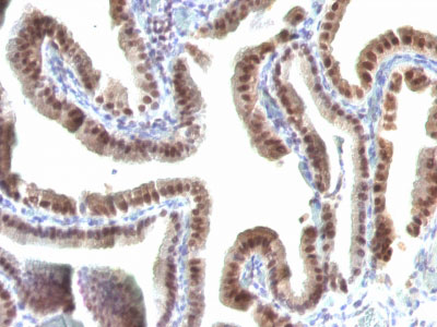
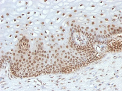
Application
| IHC, IF, FC |
|---|---|
| Primary Accession | O15405 |
| Other Accession | 27324, 460789 |
| Reactivity | Human |
| Host | Mouse |
| Clonality | Monoclonal |
| Isotype | Mouse / IgG2b, kappa |
| Clone Names | TOX3/1123 |
| Calculated MW | 63kDa |
| Gene ID | 27324 |
|---|---|
| Other Names | TOX high mobility group box family member 3, CAG trinucleotide repeat-containing gene F9 protein, Trinucleotide repeat-containing gene 9 protein, TOX3, CAGF9, TNRC9 |
| Application Note | IHC~~1:100~500 IF~~1:50~200 FC~~1:10~50 |
| Storage | Store at 2 to 8°C.Antibody is stable for 24 months. |
| Precautions | TOX3 / TNRC9 Antibody - With BSA and Azide is for research use only and not for use in diagnostic or therapeutic procedures. |
| Name | TOX3 |
|---|---|
| Synonyms | CAGF9, TNRC9 |
| Function | Transcriptional coactivator of the p300/CBP-mediated transcription complex. Activates transactivation through cAMP response element (CRE) sites. Protects against cell death by inducing antiapoptotic and repressing pro-apoptotic transcripts. Stimulates transcription from the estrogen-responsive or BCL-2 promoters. Required for depolarization-induced transcription activation of the C-FOS promoter in neurons. Associates with chromatin to the estrogen- responsive C3 promoter region. |
| Cellular Location | Nucleus. |
| Tissue Location | Expressed mainly in epithelial cells. Expressed in the central nervous system (CNS), in the ileum and within the brain in the frontal and occipital lobe. |

Thousands of laboratories across the world have published research that depended on the performance of antibodies from Abcepta to advance their research. Check out links to articles that cite our products in major peer-reviewed journals, organized by research category.
info@abcepta.com, and receive a free "I Love Antibodies" mug.

Provided below are standard protocols that you may find useful for product applications.
Background
It recognizes a 63kDa protein, which is identified as TOX3. It contains a high mobility group (HMG)-box, which regulates Ca2+-dependent transcription in neurons through interaction with the cAMP-response-element-binding protein (CREB). TOX3 appears to be associated with breast cancer susceptibility and is expressed downstream of a cytoprotective cascade together with CITED1, a transcriptional regulator that does not bind directly to DNA. TOX3 is predominantly expressed in the brain and forms homodimers. TOX3 overexpression protects neuronal cells from cell death caused by endoplasmic reticulum stress or BAX overexpression through the induction of anti-apoptotic transcripts and repression of pro-apoptotic transcripts.
References
O'Flaherty, E., et al. 2003. TOX defines a conserved subfamily of HMG-box proteins. BMC Genomics 4: 13. | Smid, M., et al. 2006. Genes associated with breast cancer metastatic to bone. J. Clin. Oncol. 24: 2261-2267. |
If you have used an Abcepta product and would like to share how it has performed, please click on the "Submit Review" button and provide the requested information. Our staff will examine and post your review and contact you if needed.
If you have any additional inquiries please email technical services at tech@abcepta.com.
Ordering Information
Other Products
Shipping Information
