


 Foundational characteristics of cancer include proliferation, angiogenesis, migration, evasion of apoptosis, and cellular immortality. Find key markers for these cellular processes and antibodies to detect them.
 The SUMOplot™ Analysis Program predicts and scores sumoylation sites in your protein. SUMOylation is a post-translational modification involved in various cellular processes, such as nuclear-cytosolic transport, transcriptional regulation, apoptosis, protein stability, response to stress, and progression through the cell cycle.
 The Autophagy Receptor Motif Plotter predicts and scores autophagy receptor binding sites in your protein. Identifying proteins connected to this pathway is critical to understanding the role of autophagy in physiological as well as pathological processes such as development, differentiation, neurodegenerative diseases, stress, infection, and cancer.
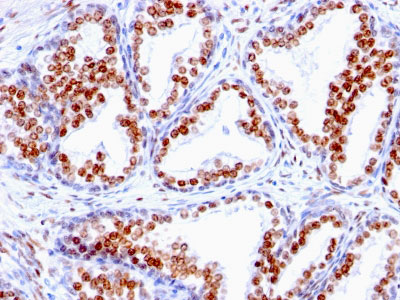
Androgen Receptor (Marker of Androgen Dependence) Antibody - With BSA and Azide
Mouse Monoclonal Antibody [Clone SPM335 ]
- SPECIFICATION
- CITATIONS
- PROTOCOLS
- BACKGROUND

Application
| IHC, IF, FC |
|---|---|
| Primary Accession | P10275 |
| Other Accession | 367, 496240 |
| Reactivity | Human |
| Host | Mouse |
| Clonality | Monoclonal |
| Isotype | Mouse / IgG1, kappa |
| Clone Names | SPM335 |
| Calculated MW | 110kDa |
| Gene ID | 367 |
|---|---|
| Other Names | Androgen receptor, Dihydrotestosterone receptor, Nuclear receptor subfamily 3 group C member 4, AR, DHTR, NR3C4 |
| Application Note | IHC~~1:100~500 IF~~1:50~200 FC~~1:10~50 |
| Storage | Store at 2 to 8°C.Antibody is stable for 24 months. |
| Precautions | Androgen Receptor (Marker of Androgen Dependence) Antibody - With BSA and Azide is for research use only and not for use in diagnostic or therapeutic procedures. |
| Name | AR |
|---|---|
| Synonyms | DHTR, NR3C4 |
| Function | Steroid hormone receptors are ligand-activated transcription factors that regulate eukaryotic gene expression and affect cellular proliferation and differentiation in target tissues (PubMed:19022849). Transcription factor activity is modulated by bound coactivator and corepressor proteins like ZBTB7A that recruits NCOR1 and NCOR2 to the androgen response elements/ARE on target genes, negatively regulating androgen receptor signaling and androgen-induced cell proliferation (PubMed:20812024). Transcription activation is also down-regulated by NR0B2. Activated, but not phosphorylated, by HIPK3 and ZIPK/DAPK3. |
| Cellular Location | Nucleus. Cytoplasm Note=Detected at the promoter of target genes (PubMed:25091737) Predominantly cytoplasmic in unligated form but translocates to the nucleus upon ligand-binding. Can also translocate to the nucleus in unligated form in the presence of RACK1. |
| Tissue Location | [Isoform 2]: Mainly expressed in heart and skeletal muscle. |

Thousands of laboratories across the world have published research that depended on the performance of antibodies from Abcepta to advance their research. Check out links to articles that cite our products in major peer-reviewed journals, organized by research category.
info@abcepta.com, and receive a free "I Love Antibodies" mug.

Provided below are standard protocols that you may find useful for product applications.
Background
Recognizes a protein of 110kDa, which is identified as androgen receptor (AR). It reacts with full length, and the newly described A form of the receptor. It does not cross react with estrogen, progesterone, or glucocorticoid receptors. The expression of AR is reportedly inversely correlated with histologic grade i.e. well differentiated prostate tumors show higher expression than the poorly differentiated tumors. In prostate cancer, AR has been proposed, as a marker of hormone-responsiveness and thus it may be useful in identifying patients likely to benefit from anti-androgen therapy. Anti-androgen receptor has been useful clinically in differentiating morpheaform basal cell carcinoma (mBCC) from desmoplastic trichoepithelioma (DTE) in the skin.ĀThis MAb is superb for staining of formalin/paraffin tissues.
References
Janne OA, et al. Androgen receptor and mechanism of androgen action. Ann Med 1993; 25:83 |
If you have used an Abcepta product and would like to share how it has performed, please click on the "Submit Review" button and provide the requested information. Our staff will examine and post your review and contact you if needed.
If you have any additional inquiries please email technical services at tech@abcepta.com.
Ordering Information
Other Products
Shipping Information
