


 Foundational characteristics of cancer include proliferation, angiogenesis, migration, evasion of apoptosis, and cellular immortality. Find key markers for these cellular processes and antibodies to detect them.
 The SUMOplot™ Analysis Program predicts and scores sumoylation sites in your protein. SUMOylation is a post-translational modification involved in various cellular processes, such as nuclear-cytosolic transport, transcriptional regulation, apoptosis, protein stability, response to stress, and progression through the cell cycle.
 The Autophagy Receptor Motif Plotter predicts and scores autophagy receptor binding sites in your protein. Identifying proteins connected to this pathway is critical to understanding the role of autophagy in physiological as well as pathological processes such as development, differentiation, neurodegenerative diseases, stress, infection, and cancer.
CD45RA (Leucocyte Marker) Antibody - With BSA and Azide
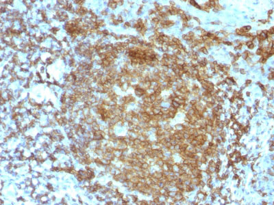
Mouse Monoclonal Antibody [Clone PTPRC/818 ]
- SPECIFICATION
- CITATIONS
- PROTOCOLS
- BACKGROUND

Application
| IHC, IF, FC |
|---|---|
| Primary Accession | P08575 |
| Other Accession | 5788, 654514 |
| Reactivity | Human |
| Host | Mouse |
| Clonality | Monoclonal |
| Isotype | Mouse / IgG2a, kappa |
| Clone Names | PTPRC/818 |
| Calculated MW | 205-220kDa |
| Gene ID | 5788 |
|---|---|
| Other Names | Receptor-type tyrosine-protein phosphatase C, 3.1.3.48, Leukocyte common antigen, L-CA, T200, CD45, PTPRC, CD45 |
| Application Note | IHC~~1:100~500 IF~~1:50~200 FC~~1:10~50 |
| Storage | Store at 2 to 8°C.Antibody is stable for 24 months. |
| Precautions | CD45RA (Leucocyte Marker) Antibody - With BSA and Azide is for research use only and not for use in diagnostic or therapeutic procedures. |
| Name | PTPRC (HGNC:9666) |
|---|---|
| Synonyms | CD45 |
| Function | Protein tyrosine-protein phosphatase required for T-cell activation through the antigen receptor (PubMed:35767951). Acts as a positive regulator of T-cell coactivation upon binding to DPP4. The first PTPase domain has enzymatic activity, while the second one seems to affect the substrate specificity of the first one. Upon T-cell activation, recruits and dephosphorylates SKAP1 and FYN. Dephosphorylates LYN, and thereby modulates LYN activity (By similarity). Interacts with CLEC10A at antigen presenting cell-T cell contact; CLEC10A on immature dendritic cells recognizes Tn antigen- carrying PTPRC/CD45 receptor on effector T cells and modulates T cell activation threshold to limit autoreactivity. |
| Cellular Location | Cell membrane; Single-pass type I membrane protein. Membrane raft. Synapse. Note=Colocalized with DPP4 in membrane rafts. |
| Tissue Location | Isoform 1: Detected in thymocytes. Isoform 2: Detected in thymocytes. Isoform 3: Detected in thymocytes. Isoform 4: Not detected in thymocytes. Isoform 5: Detected in thymocytes. Isoform 6: Not detected in thymocytes. Isoform 7: Detected in thymocytes Isoform 8: Not detected in thymocytes. |

Thousands of laboratories across the world have published research that depended on the performance of antibodies from Abcepta to advance their research. Check out links to articles that cite our products in major peer-reviewed journals, organized by research category.
info@abcepta.com, and receive a free "I Love Antibodies" mug.

Provided below are standard protocols that you may find useful for product applications.
Background
Recognizes a protein of 205kDa-220kDa, identified as CD45RA. CD45RA is isoforms of the human leukocyte common antigen (CD45). Human CD45 contains three exons which encode peptide segments designated A, B and C, respectively. The differential splicing of the exons generates at least five isoforms, ABC, AB, BC, B and O. This antibody reacts with ABC and BC isoforms. CD45RA is expressed on 40-50% of peripheral CD4+ T-cells, 50% of peripheral CD8+ T-cells, B-cells, and leukemic B-cell lines. T-cells expressing CD45RA are naive or virgin T-cells. T-cells expressing CD45RO are memory T-cells. CD45RA and CD45RO define complementary, predominantly non-overlapping populations of resting peripheral T-cells. This MAb is useful in study on the subpopulation of CD4+ or CD8+ T-cells. It can especially be used to differentiate T-cell lymphomas (CD45RO +ve) from B cell lymphomas (CD45RA +ve).
References
West, K.P., et al. 1986. The demonstration of B-cell, T-cell and myeloid antigens in paraffin sections. J. Pathol. 150: 89-101. | Streuli, M., et al. 1987. Differential usage of three exons generates at least five different mRNAs encoding human leukocyte common antigens. J. Exp. Med. 166: 1548-1566
If you have used an Abcepta product and would like to share how it has performed, please click on the "Submit Review" button and provide the requested information. Our staff will examine and post your review and contact you if needed.
If you have any additional inquiries please email technical services at tech@abcepta.com.

Ordering Information
Other Products
Shipping Information
