


 Foundational characteristics of cancer include proliferation, angiogenesis, migration, evasion of apoptosis, and cellular immortality. Find key markers for these cellular processes and antibodies to detect them.
 The SUMOplot™ Analysis Program predicts and scores sumoylation sites in your protein. SUMOylation is a post-translational modification involved in various cellular processes, such as nuclear-cytosolic transport, transcriptional regulation, apoptosis, protein stability, response to stress, and progression through the cell cycle.
 The Autophagy Receptor Motif Plotter predicts and scores autophagy receptor binding sites in your protein. Identifying proteins connected to this pathway is critical to understanding the role of autophagy in physiological as well as pathological processes such as development, differentiation, neurodegenerative diseases, stress, infection, and cancer.
Cytokeratin 10/13 Antibody - With BSA and Azide
Mouse Monoclonal Antibody [Clone SPM262 ]
- SPECIFICATION
- CITATIONS
- PROTOCOLS
- BACKGROUND

Application
| WB, IHC-P, IF, FC |
|---|---|
| Primary Accession | P13645 |
| Other Accession | 3858, 99936 |
| Reactivity | Human, Cat |
| Host | Mouse |
| Clonality | Monoclonal |
| Isotype | Mouse / IgG2a, kappa |
| Clone Names | SPM262 |
| Calculated MW | 56.5kDa (CK10) & 53kDa (CK13) |
| Gene ID | 3858 |
|---|---|
| Other Names | Keratin, type I cytoskeletal 10, Cytokeratin-10, CK-10, Keratin-10, K10, KRT10, KPP |
| Application Note | WB~~1:1000 IHC-P~~N/A IF~~1:50~200 FC~~1:10~50 |
| Storage | Store at 2 to 8°C.Antibody is stable for 24 months. |
| Precautions | Cytokeratin 10/13 Antibody - With BSA and Azide is for research use only and not for use in diagnostic or therapeutic procedures. |
| Name | KRT10 |
|---|---|
| Synonyms | KPP |
| Function | Plays a role in the establishment of the epidermal barrier on plantar skin (By similarity). Involved in the maintenance of cell layer development and keratin filament bundles in suprabasal cells of the epithelium (By similarity). |
| Cellular Location | Secreted, extracellular space. Cell surface. Cytoplasm |
| Tissue Location | Seen in all suprabasal cell layers including stratum corneum. Expressed on the surface of lung cell lines (PubMed:19627498). Localized on the surface of desquamated nasal epithelial cells (at protein level) (PubMed:12427098) |

Thousands of laboratories across the world have published research that depended on the performance of antibodies from Abcepta to advance their research. Check out links to articles that cite our products in major peer-reviewed journals, organized by research category.
info@abcepta.com, and receive a free "I Love Antibodies" mug.

Provided below are standard protocols that you may find useful for product applications.
Background
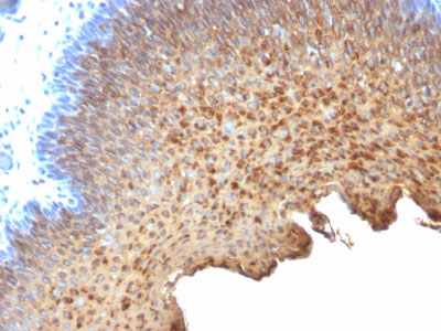
This antibody recognizes cytokeratin 10 (56.5kDa) and cytokeratin 13 (53kDa) in Western blotting. It recognizes only cytokeratin 13 in formalin-fixed, paraffin-embedded tissue sections. It does not react with cytokeratin 10 positive, cytokeratin 13 negative epithelia such as epidermis. However, on frozen sections this MAb serves as differentiation-related marker of all stratified epithelia; it stains all suprabasal cells in both cornifying and non-cornifying stratified epithelia and more differentiated cells of squamous carcinomas.
References
Ivanyi D et. al. Journal of Pathology, 1989, 159:7-12.,Ivanyi, D., Minke, J. M., Hageman, C., Groeneveld, E., and van Doornewaard, G. (1992). Patterns of expression of feline cytokeratins in healthy epithelia and mammary carcinoma cells, Am J Vet Res 53, 304-14. ,Ivanyi, D., Minke, J. M., Hageman, C., Groeneveld, E., van Doornewaard, G., and Misdorp, W. (1993). Cytokeratins as markers of initial stages of squamous metaplasia in feline mammary carcinomas, Am J Vet Res 54, 1095
If you have used an Abcepta product and would like to share how it has performed, please click on the "Submit Review" button and provide the requested information. Our staff will examine and post your review and contact you if needed.
If you have any additional inquiries please email technical services at tech@abcepta.com.
Ordering Information
Other Products
Shipping Information
