


 Foundational characteristics of cancer include proliferation, angiogenesis, migration, evasion of apoptosis, and cellular immortality. Find key markers for these cellular processes and antibodies to detect them.
 The SUMOplot™ Analysis Program predicts and scores sumoylation sites in your protein. SUMOylation is a post-translational modification involved in various cellular processes, such as nuclear-cytosolic transport, transcriptional regulation, apoptosis, protein stability, response to stress, and progression through the cell cycle.
 The Autophagy Receptor Motif Plotter predicts and scores autophagy receptor binding sites in your protein. Identifying proteins connected to this pathway is critical to understanding the role of autophagy in physiological as well as pathological processes such as development, differentiation, neurodegenerative diseases, stress, infection, and cancer.
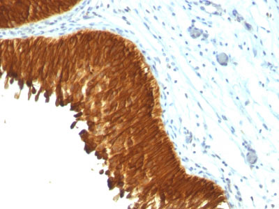
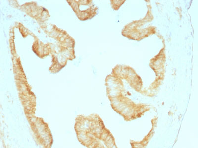
Cytokeratin, Multi (Epithelial Marker) Antibody - With BSA and Azide
Mouse Monoclonal Antibody [Clone KRT/457 ]
- SPECIFICATION
- CITATIONS
- PROTOCOLS
- BACKGROUND

Application
| WB, IHC-P, IF, FC |
|---|---|
| Primary Accession | P02538 |
| Other Accession | 3851 (CK4), 3852 (CK5), 3853 (CK6A), 3854 (CK6B), 286887 (CK6C), 3856 (CK8), 3858 (CK10), 3860 (CK13), 3875 (CK18) (Human), P04259 (CK6B), P13647 (CK5), P19013 (CK4), P48668 (CK6C) |
| Reactivity | Human, Mouse, Rat, Monkey, Pig, Goat, Bovine, Guinea Pig |
| Host | Mouse |
| Clonality | Monoclonal |
| Isotype | Mouse / IgG1 |
| Clone Names | KRT/457 |
| Calculated MW | Multiple KDa |
| Gene ID | 3853 |
|---|---|
| Other Names | Keratin, type II cytoskeletal 6A, Cytokeratin-6A, CK-6A, Cytokeratin-6D, CK-6D, Keratin-6A, K6A, Type-II keratin Kb6, Hom s 5, KRT6A, K6A, KRT6D |
| Application Note | WB~~1:1000 IHC-P~~N/A IF~~1:50~200 FC~~1:10~50 |
| Storage | Store at 2 to 8°C.Antibody is stable for 24 months. |
| Precautions | Cytokeratin, Multi (Epithelial Marker) Antibody - With BSA and Azide is for research use only and not for use in diagnostic or therapeutic procedures. |
| Name | KRT6A |
|---|---|
| Synonyms | K6A, KRT6D |
| Function | Epidermis-specific type I keratin involved in wound healing. Involved in the activation of follicular keratinocytes after wounding, while it does not play a major role in keratinocyte proliferation or migration. Participates in the regulation of epithelial migration by inhibiting the activity of SRC during wound repair. |
| Tissue Location | Expressed in the corneal epithelium (at protein level). |

Thousands of laboratories across the world have published research that depended on the performance of antibodies from Abcepta to advance their research. Check out links to articles that cite our products in major peer-reviewed journals, organized by research category.
info@abcepta.com, and receive a free "I Love Antibodies" mug.

Provided below are standard protocols that you may find useful for product applications.
Background
Twenty human keratins are resolved with two-dimensional gel electrophoresis into acidic (pI 6.0) subfamilies. This antibody recognizes acidic (Type I or LMW) and basic (Type II or HMW) cytokeratins, including 59kDa (CK4); 58kDa (CK5); 56kDa (CK6); 52kDa (CK8); 56.5kDa (CK10); 53kDa (CK13) and 45kDa (CK18). This is a broad-spectrum antibody, which has been reported to differentiate epithelial tumors from non-epithelial tumors. Many studies have shown the usefulness of keratins as markers in cancer research and tumor diagnosis.
References
Bartek J et. al. J Pathol, 1991, 164(3):215-24. ,Kasper M. Histochemistry, 1991, 95(6):613-20
If you have used an Abcepta product and would like to share how it has performed, please click on the "Submit Review" button and provide the requested information. Our staff will examine and post your review and contact you if needed.
If you have any additional inquiries please email technical services at tech@abcepta.com.
Ordering Information
Other Products
Shipping Information
