


 Foundational characteristics of cancer include proliferation, angiogenesis, migration, evasion of apoptosis, and cellular immortality. Find key markers for these cellular processes and antibodies to detect them.
 The SUMOplot™ Analysis Program predicts and scores sumoylation sites in your protein. SUMOylation is a post-translational modification involved in various cellular processes, such as nuclear-cytosolic transport, transcriptional regulation, apoptosis, protein stability, response to stress, and progression through the cell cycle.
 The Autophagy Receptor Motif Plotter predicts and scores autophagy receptor binding sites in your protein. Identifying proteins connected to this pathway is critical to understanding the role of autophagy in physiological as well as pathological processes such as development, differentiation, neurodegenerative diseases, stress, infection, and cancer.
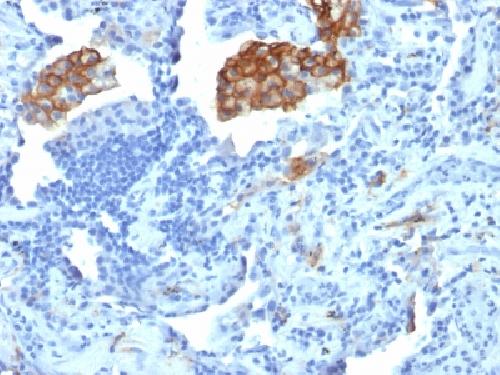
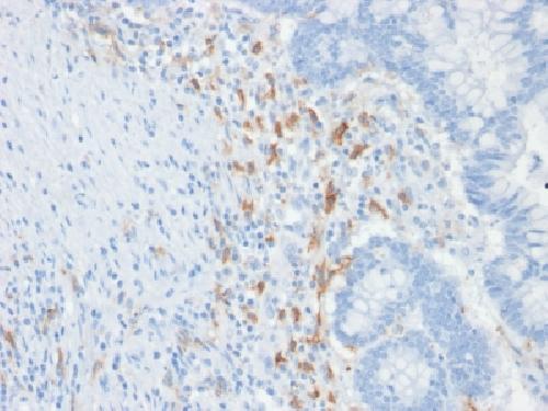
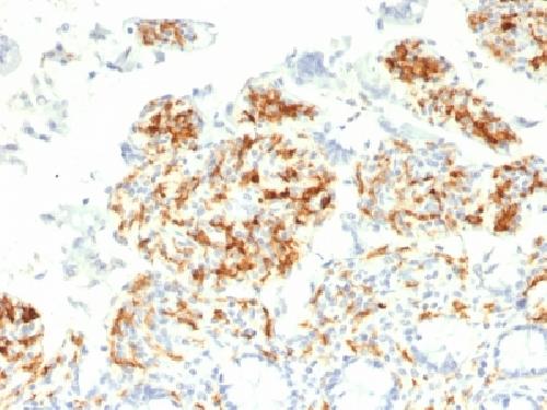
Anti-CD209 / DC-SIGN Antibody
Mouse Monoclonal Antibody
- SPECIFICATION
- CITATIONS
- PROTOCOLS
- BACKGROUND

Application
| IHC-P, IF, FC |
|---|---|
| Primary Accession | Q9NNX6 |
| Other Accession | 278694 |
| Reactivity | Human |
| Host | Mouse |
| Clonality | Monoclonal |
| Isotype | Mouse / IgG2b, kappa |
| Clone Names | C209/1781 |
| Calculated MW | 45775 Da |
| Gene ID | 30835 |
|---|---|
| Other Names | CD209; CDSIGN; CIRE; CLEC4L; DC-SIGN; DC-SIGN1; DCSIGN; Dendritic cell-specific ICAM-3 Grabbing Non-integrin 1; HIV GP120 Binding Protein; SIGN-R1; SIGNR5 |
| Application Note | Flow Cytometry (0.5-1ug/million cells); Immunofluorescence (1-2ug/ml); ,Immunohistology (Formalin-fixed) (0.5-1ug/ml for 30 minutes at RT),(Staining of formalin-fixed tissues is enhanced by boiling tissue sections in 10mM Tris with 1mM EDTA Buffer, pH 9.0, for 10-20 min followed by cooling at RT for 20 minutes),Optimal dilution for a specific application should be determined. |
| Format | 200ug/ml of Ab purified from Bioreactor Concentrate by Protein A/G. Prepared in 10mM PBS with 0.05% BSA & 0.05% azide. Also available WITHOUT BSA & azide at 1.0mg/ml. |
| Storage | Store at 2 to 8°C.Antibody is stable for 24 months. |
| Precautions | Anti-CD209 / DC-SIGN Antibody is for research use only and not for use in diagnostic or therapeutic procedures. |
| Name | CD209 |
|---|---|
| Synonyms | CLEC4L |
| Function | Pathogen-recognition receptor expressed on the surface of immature dendritic cells (DCs) and involved in initiation of primary immune response. Thought to mediate the endocytosis of pathogens which are subsequently degraded in lysosomal compartments. The receptor returns to the cell membrane surface and the pathogen-derived antigens are presented to resting T-cells via MHC class II proteins to initiate the adaptive immune response. |
| Cellular Location | [Isoform 1]: Cell membrane; Single- pass type II membrane protein [Isoform 3]: Cell membrane; Single- pass type II membrane protein [Isoform 5]: Cell membrane; Single- pass type II membrane protein [Isoform 7]: Secreted. [Isoform 9]: Secreted. [Isoform 11]: Secreted. |
| Tissue Location | Predominantly expressed in dendritic cells and in DC-residing tissues. Also found in placental macrophages, endothelial cells of placental vascular channels, peripheral blood mononuclear cells, and THP-1 monocytes. |

Thousands of laboratories across the world have published research that depended on the performance of antibodies from Abcepta to advance their research. Check out links to articles that cite our products in major peer-reviewed journals, organized by research category.
info@abcepta.com, and receive a free "I Love Antibodies" mug.

Provided below are standard protocols that you may find useful for product applications.
Background
DC-SIGN is a transmembrane receptor that is expressed on the surface of dendritic cells and macrophages. It is involved in the innate immune system and recognizes numerous evolutionarily divergent pathogens ranging from parasites to viruses. The protein is organized into three distinct domains: an N-terminal transmembrane domain, a tandem-repeat neck domain and C-type lectin carbohydrate recognition domain. The extracellular region consisting of the C-type lectin and neck domains has a dual function as a pathogen recognition receptor and a cell adhesion receptor by binding carbohydrate ligands on the surface of microbes and endogenous cells. The neck region is important for homo-oligomerization, which allows the receptor to bind multivalent ligands with high avidity.
If you have used an Abcepta product and would like to share how it has performed, please click on the "Submit Review" button and provide the requested information. Our staff will examine and post your review and contact you if needed.
If you have any additional inquiries please email technical services at tech@abcepta.com.
Ordering Information
Other Products
Shipping Information
