


 Foundational characteristics of cancer include proliferation, angiogenesis, migration, evasion of apoptosis, and cellular immortality. Find key markers for these cellular processes and antibodies to detect them.
 The SUMOplot™ Analysis Program predicts and scores sumoylation sites in your protein. SUMOylation is a post-translational modification involved in various cellular processes, such as nuclear-cytosolic transport, transcriptional regulation, apoptosis, protein stability, response to stress, and progression through the cell cycle.
 The Autophagy Receptor Motif Plotter predicts and scores autophagy receptor binding sites in your protein. Identifying proteins connected to this pathway is critical to understanding the role of autophagy in physiological as well as pathological processes such as development, differentiation, neurodegenerative diseases, stress, infection, and cancer.
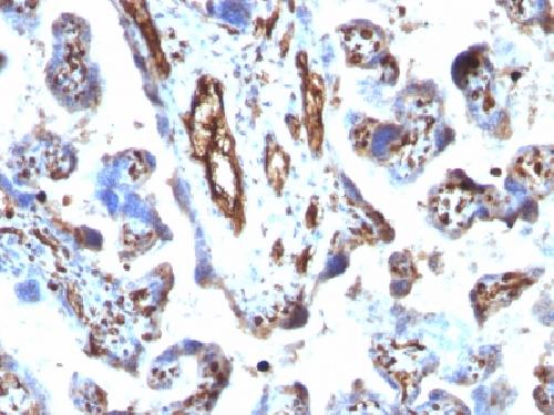
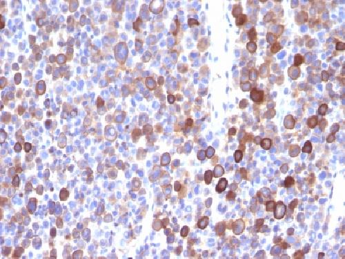
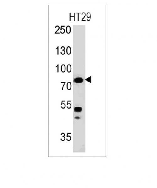
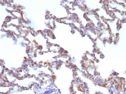
Anti-Moesin Antibody
Mouse Monoclonal Antibody
- SPECIFICATION
- CITATIONS
- PROTOCOLS
- BACKGROUND

Application
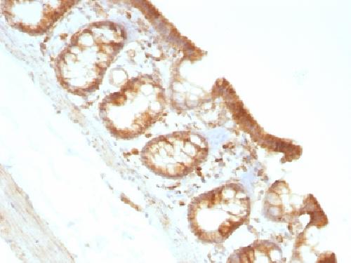
| WB, IHC-P, IF, FC |
|---|---|
| Primary Accession | P26038 |
| Other Accession | 87752 |
| Reactivity | Human, Rat |
| Host | Mouse |
| Clonality | Monoclonal |
| Isotype | Mouse / IgG1, kappa |
| Clone Names | MSN/491 |
| Calculated MW | 67820 Da |
| Gene ID | 4478 |
|---|---|
| Other Names | Membrane-organizing extension spike protein; Moesin/anaplastic lymphoma kinase fusion protein; MSN/ALK fusion |
| Application Note | Flow Cytometry (0.5-1ug/million cells); Immunofluorescence (0.5-1ug/ml); ,Western Blotting (0.5-1ug/ml); ,Immunohistology (Formalin-fixed) (0.5-1ug/ml for 30 minutes at RT),(Staining of formalin-fixed tissues requires boiling tissue sections in 10mM citrate buffer, pH 6.0, for 10-20 min followed by cooling at RT for 20 minutes),Optimal dilution for a specific application should be determined. |
| Format | 200ug/ml of Ab purified from Bioreactor Concentrate by Protein A/G. Prepared in 10mM PBS with 0.05% BSA & 0.05% azide. Also available WITHOUT BSA & azide at 1.0mg/ml. |
| Storage | Store at 2 to 8°C.Antibody is stable for 24 months. |
| Precautions | Anti-Moesin Antibody is for research use only and not for use in diagnostic or therapeutic procedures. |
| Name | MSN (HGNC:7373) |
|---|---|
| Function | Ezrin-radixin-moesin (ERM) family protein that connects the actin cytoskeleton to the plasma membrane and thereby regulates the structure and function of specific domains of the cell cortex. Tethers actin filaments by oscillating between a resting and an activated state providing transient interactions between moesin and the actin cytoskeleton (PubMed:10212266). Once phosphorylated on its C-terminal threonine, moesin is activated leading to interaction with F-actin and cytoskeletal rearrangement (PubMed:10212266). These rearrangements regulate many cellular processes, including cell shape determination, membrane transport, and signal transduction (PubMed:12387735, PubMed:15039356). The role of moesin is particularly important in immunity acting on both T and B-cells homeostasis and self-tolerance, regulating lymphocyte egress from lymphoid organs (PubMed:9298994, PubMed:9616160). Modulates phagolysosomal biogenesis in macrophages (By similarity). Also participates in immunologic synapse formation (PubMed:27405666). |
| Cellular Location | Cell membrane; Peripheral membrane protein {ECO:0000250|UniProtKB:P26041}; Cytoplasmic side {ECO:0000250|UniProtKB:P26041}. Cytoplasm, cytoskeleton {ECO:0000250|UniProtKB:P26041}. Apical cell membrane {ECO:0000250|UniProtKB:P26041}; Peripheral membrane protein {ECO:0000250|UniProtKB:P26041}; Cytoplasmic side {ECO:0000250|UniProtKB:P26041}. Cell projection, microvillus membrane {ECO:0000250|UniProtKB:P26041}; Peripheral membrane protein {ECO:0000250|UniProtKB:P26041}; Cytoplasmic side {ECO:0000250|UniProtKB:P26041}. Cell projection, microvillus {ECO:0000250|UniProtKB:P26041}. Note=Phosphorylated form is enriched in microvilli-like structures at apical membrane. Increased cell membrane localization of both phosphorylated and non-phosphorylated forms seen after thrombin treatment (By similarity). Localizes at the uropods of T lymphoblasts. {ECO:0000250|UniProtKB:P26041, ECO:0000269|PubMed:18586956, ECO:0000269|PubMed:9298994} |
| Tissue Location | In all tissues and cultured cells studied. |

Thousands of laboratories across the world have published research that depended on the performance of antibodies from Abcepta to advance their research. Check out links to articles that cite our products in major peer-reviewed journals, organized by research category.
info@abcepta.com, and receive a free "I Love Antibodies" mug.

Provided below are standard protocols that you may find useful for product applications.
Background
Recognizes 78kDa moesin protein. Moesin, a member of the talin-4.1 superfamily, is a linking protein of the sub-membranous actin cytoskeleton. It is expressed in variable amounts in cells of different phenotypes such as macrophages, lymphocytes, fibroblastic, endothelial, epithelial, and neuronal cell lines but not in blood cells. The ERM proteins, ezrin, radixin, and moesin are involved in a variety of cellular functions, such as cell adhesion, migration, and the organization of cell surface structures, and are highly homologous, both in protein sequence and in functional activity, with merlin/schwannomin, a neurofibromatosis-2-associated tumor-suppressor protein. Cell lines of epithelial and mesothelial origin contain both moesin and radixin whereas cells of endothelial and lymphoid origin express moesin.
If you have used an Abcepta product and would like to share how it has performed, please click on the "Submit Review" button and provide the requested information. Our staff will examine and post your review and contact you if needed.
If you have any additional inquiries please email technical services at tech@abcepta.com.
Ordering Information
Other Products
Shipping Information
