


 Foundational characteristics of cancer include proliferation, angiogenesis, migration, evasion of apoptosis, and cellular immortality. Find key markers for these cellular processes and antibodies to detect them.
 The SUMOplot™ Analysis Program predicts and scores sumoylation sites in your protein. SUMOylation is a post-translational modification involved in various cellular processes, such as nuclear-cytosolic transport, transcriptional regulation, apoptosis, protein stability, response to stress, and progression through the cell cycle.
 The Autophagy Receptor Motif Plotter predicts and scores autophagy receptor binding sites in your protein. Identifying proteins connected to this pathway is critical to understanding the role of autophagy in physiological as well as pathological processes such as development, differentiation, neurodegenerative diseases, stress, infection, and cancer.
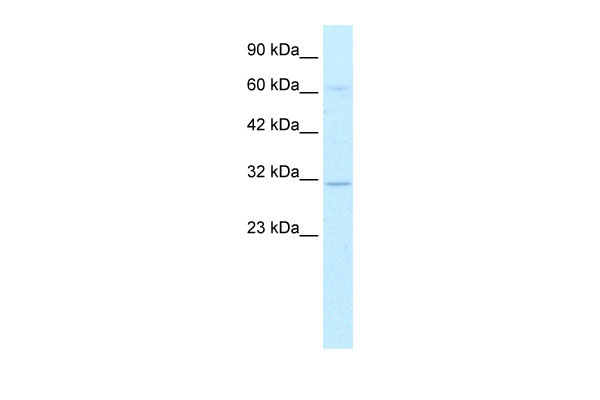
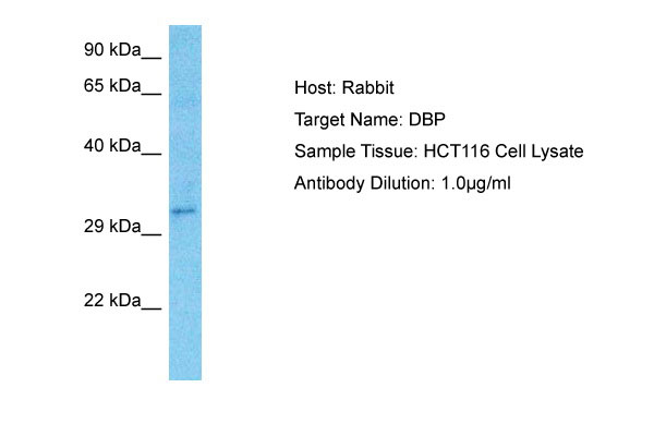
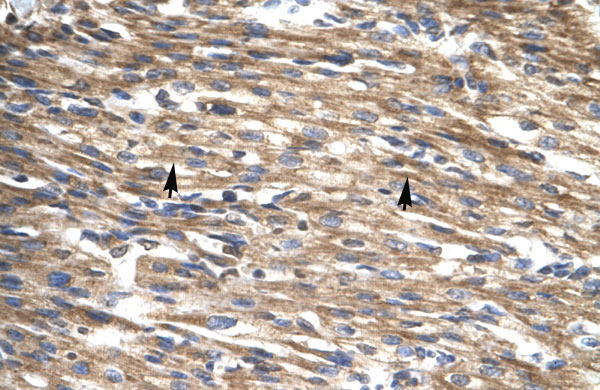
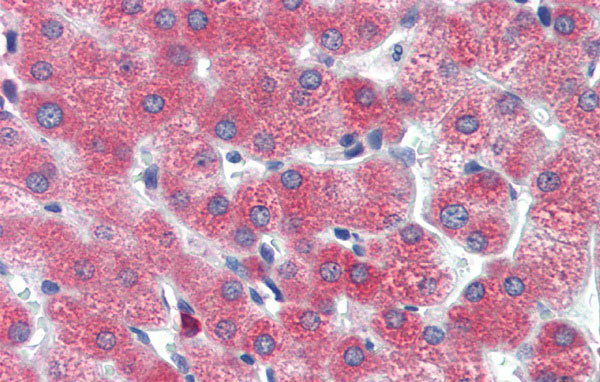
DBP antibody - middle region
Rabbit Polyclonal Antibody
- SPECIFICATION
- CITATIONS: 2
- PROTOCOLS
- BACKGROUND

Application
| WB, IHC |
|---|---|
| Primary Accession | Q10586 |
| Other Accession | NM_001352, NP_001343 |
| Reactivity | Human, Mouse, Rat, Pig, Sheep, Horse, Bovine, Dog |
| Predicted | Human, Mouse, Rat, Pig, Sheep, Bovine, Dog |
| Host | Rabbit |
| Clonality | Polyclonal |
| Calculated MW | 34kDa |
| Gene ID | 1628 |
|---|---|
| Alias Symbol | DABP |
| Other Names | D site-binding protein, Albumin D box-binding protein, Albumin D-element-binding protein, Tax-responsive enhancer element-binding protein 302, TaxREB302, DBP |
| Format | Liquid. Purified antibody supplied in 1x PBS buffer with 0.09% (w/v) sodium azide and 2% sucrose. |
| Reconstitution & Storage | Add 50 ul of distilled water. Final anti-DBP antibody concentration is 1 mg/ml in PBS buffer with 2% sucrose. For longer periods of storage, store at 20°C. Avoid repeat freeze-thaw cycles. |
| Precautions | DBP antibody - middle region is for research use only and not for use in diagnostic or therapeutic procedures. |
| Name | DBP |
|---|---|
| Function | This transcriptional activator recognizes and binds to the sequence 5'-RTTAYGTAAY-3' found in the promoter of genes such as albumin, CYP2A4 and CYP2A5. It is not essential for circadian rhythm generation, but modulates important clock output genes. May be a direct target for regulation by the circadian pacemaker component clock. May affect circadian period and sleep regulation. |
| Cellular Location | Nucleus. |
| Tissue Location | Ubiquitously expressed. Expressed in the suprachiasmatic nuclei (SCN) and in most peripheral tissues, with a strong circadian rhythmicity |
Citations ( 0 )

Application Protocols
Provided below are standard protocols that you may find useful for product applications.
References
Smith,J.S., et al., (2000) Genomics 64 (1), 44-50Reconstitution and Storage:For short term use, store at 2-8C up to 1 week. For long term storage, store at -20C in small aliquots to prevent freeze-thaw cycles.
Abcepta welcomes feedback from its customers.
If you have used an Abcepta product and would like to share how it has performed, please click on the "Submit Review" button and provide the requested information. Our staff will examine and post your review and contact you if needed.
If you have any additional inquiries please email technical services at tech@abcepta.com.
$ 389.00
Cat# AI10247
Ordering Information
United States
AlbaniaAustraliaAustriaBelgiumBosnia & HerzegovinaBrazilBulgariaCanadaCentral AmericaChinaCroatiaCyprusCzech RepublicDenmarkEstoniaFinlandFranceGermanyGreeceHong KongHungaryIcelandIndiaIndonesiaIrelandIsraelItalyJapanLatviaLithuaniaLuxembourgMacedoniaMalaysiaMaltaMexicoNetherlandsNew ZealandNorwayPakistanPolandPortugalRomaniaSerbiaSingaporeSlovakiaSloveniaSouth AfricaSouth KoreaSpainSwedenSwitzerlandTaiwanTurkeyUnited KingdomUnited StatesVietnamWorldwideOthers
USA Headquarters
(888) 735-7227 / (858) 622-0099 or (858) 875-1900
Other Products
Shipping Information
Domestic orders (in stock items)
Shipped out the same day. Orders placed after 1 PM (PST) will ship out the next business day.
International orders
Contact your local distributors
