


 Foundational characteristics of cancer include proliferation, angiogenesis, migration, evasion of apoptosis, and cellular immortality. Find key markers for these cellular processes and antibodies to detect them.
 The SUMOplot™ Analysis Program predicts and scores sumoylation sites in your protein. SUMOylation is a post-translational modification involved in various cellular processes, such as nuclear-cytosolic transport, transcriptional regulation, apoptosis, protein stability, response to stress, and progression through the cell cycle.
 The Autophagy Receptor Motif Plotter predicts and scores autophagy receptor binding sites in your protein. Identifying proteins connected to this pathway is critical to understanding the role of autophagy in physiological as well as pathological processes such as development, differentiation, neurodegenerative diseases, stress, infection, and cancer.
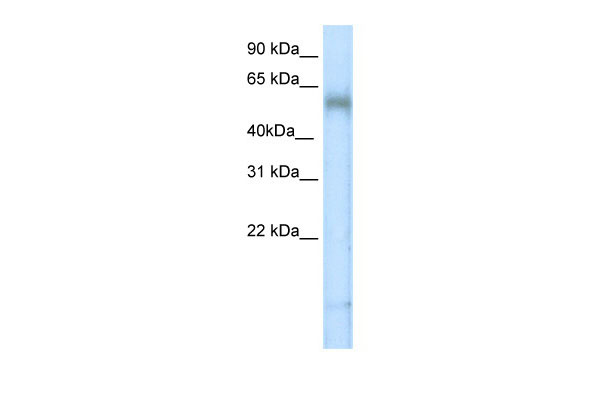
TBX6 antibody - N-terminal region
Rabbit Polyclonal Antibody
- SPECIFICATION
- CITATIONS
- PROTOCOLS
- BACKGROUND

Application
| WB |
|---|---|
| Primary Accession | O95947 |
| Other Accession | NM_004608, NP_004599 |
| Reactivity | Human, Mouse, Rat, Pig, Horse, Bovine, Dog |
| Predicted | Human, Mouse, Rat, Bovine, Dog |
| Host | Rabbit |
| Clonality | Polyclonal |
| Calculated MW | 47kDa |
| Gene ID | 6911 |
|---|---|
| Other Names | T-box transcription factor TBX6, T-box protein 6, TBX6 |
| Format | Liquid. Purified antibody supplied in 1x PBS buffer with 0.09% (w/v) sodium azide and 2% sucrose. |
| Reconstitution & Storage | Add 100 ul of distilled water. Final anti-TBX6 antibody concentration is 1 mg/ml in PBS buffer with 2% sucrose. For longer periods of storage, store at 20°C. Avoid repeat freeze-thaw cycles. |
| Precautions | TBX6 antibody - N-terminal region is for research use only and not for use in diagnostic or therapeutic procedures. |
| Name | TBX6 |
|---|---|
| Function | T-box transcription factor that plays an essential role in the determination of the fate of axial stem cells: neural vs mesodermal. Acts in part by down-regulating, a specific enhancer (N1) of SOX2, to inhibit neural development. Seems to play also an essential role in left/right axis determination and acts through effects on Notch signaling around the node as well as through an effect on the morphology and motility of the nodal cilia (By similarity). |
| Cellular Location | Nucleus {ECO:0000255|PROSITE-ProRule:PRU00201}. |
| Tissue Location | Expressed in fetal tail bud, posterior spinal tissue, intervertebral disk and testis. Also expressed in adult testis, kidney, lung, muscle and thymus |

Thousands of laboratories across the world have published research that depended on the performance of antibodies from Abcepta to advance their research. Check out links to articles that cite our products in major peer-reviewed journals, organized by research category.
info@abcepta.com, and receive a free "I Love Antibodies" mug.

Provided below are standard protocols that you may find useful for product applications.
References
Papapetrou,C., et al., (1999) Genomics 55 (2), 238-241Reconstitution and Storage:For short term use, store at 2-8C up to 1 week. For long term storage, store at -20C in small aliquots to prevent freeze-thaw cycles.
If you have used an Abcepta product and would like to share how it has performed, please click on the "Submit Review" button and provide the requested information. Our staff will examine and post your review and contact you if needed.
If you have any additional inquiries please email technical services at tech@abcepta.com.
Ordering Information
Other Products
Shipping Information
