


 Foundational characteristics of cancer include proliferation, angiogenesis, migration, evasion of apoptosis, and cellular immortality. Find key markers for these cellular processes and antibodies to detect them.
 The SUMOplot™ Analysis Program predicts and scores sumoylation sites in your protein. SUMOylation is a post-translational modification involved in various cellular processes, such as nuclear-cytosolic transport, transcriptional regulation, apoptosis, protein stability, response to stress, and progression through the cell cycle.
 The Autophagy Receptor Motif Plotter predicts and scores autophagy receptor binding sites in your protein. Identifying proteins connected to this pathway is critical to understanding the role of autophagy in physiological as well as pathological processes such as development, differentiation, neurodegenerative diseases, stress, infection, and cancer.
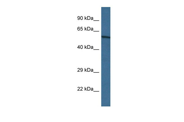
Accn2 antibody - N-terminal region
Rabbit Polyclonal Antibody
- SPECIFICATION
- CITATIONS
- PROTOCOLS
- BACKGROUND

Application
| WB |
|---|---|
| Primary Accession | Q6NXK8 |
| Other Accession | NM_009597, NP_033727 |
| Reactivity | Human, Mouse, Rat, Rabbit, Horse, Bovine, Dog |
| Predicted | Human, Mouse, Rat, Rabbit, Bovine |
| Host | Rabbit |
| Clonality | Polyclonal |
| Calculated MW | 60kDa |
| Gene ID | 11419 |
|---|---|
| Alias Symbol | AI843610, ASIC, ASIC1, ASIC1a, B530003N02Rik, BNaC2, Accn2 |
| Other Names | Acid-sensing ion channel 1, ASIC1, Acid-sensing ion channel, Amiloride-sensitive cation channel 2, neuronal, Brain sodium channel 2, BNaC2, Asic1, Accn2, Asic, Bnac2 |
| Format | Liquid. Purified antibody supplied in 1x PBS buffer with 0.09% (w/v) sodium azide and 2% sucrose. |
| Reconstitution & Storage | Add 50 ul of distilled water. Final anti-Accn2 antibody concentration is 1 mg/ml in PBS buffer with 2% sucrose. For longer periods of storage, store at 20°C. Avoid repeat freeze-thaw cycles. |
| Precautions | Accn2 antibody - N-terminal region is for research use only and not for use in diagnostic or therapeutic procedures. |
| Name | Asic1 {ECO:0000312|MGI:MGI:1194915} |
|---|---|
| Function | Forms voltage-independent, pH-gated trimeric sodium channels that act as postsynaptic excitatory receptors in the nervous system, playing a crucial role in regulating synaptic plasticity, learning, and memory (PubMed:11988176, PubMed:12843249, PubMed:15369669, PubMed:17060608). Upon extracellular pH drop this channel elicits transient, fast activating, and completely desensitizing inward currents (PubMed:15369669). Displays high selectivity for sodium ions but can also permit the permeation of other cations (PubMed:15369669). Regulates more or less directly intracellular calcium concentration and CaMKII phosphorylation, and thereby the density of dendritic spines (PubMed:15369669, PubMed:17060608). Modulates neuronal activity in the circuits underlying innate fear (PubMed:17662962). |
| Cellular Location | Cell membrane; Multi-pass membrane protein {ECO:0000250|UniProtKB:P78348} Postsynaptic cell membrane. Cell projection, dendrite. Note=Isolated in synaptosomes from the dendritic synapses of neurons. |
| Tissue Location | Expressed in brain areas receiving strong excitatory corticofugal input. In hippocampus, expressed in the hilus of the dentate gyrus. In the cerebral cortex expressed in anterior and posterior cingulate cortex, sensory and motor cortices. In the sensory cortex strongest expression is detected in the whisker barrel field. In sensorimotor and cingulate cortex expression is elevated in layer III Also expressed in basal ganglia, striatum, ventral pallidum, olfactory tubercle, and nucleus accumbens. Weakly expressed in thalamus with the exception of the habenula and the medial septal nuclei. In olfactory bulb, preferentially expressed in the glomerular layer, within glomeruli. Expressed in cerebellum in the molecular and granule cell layers. Strongly expressed in amygdala complex, particularly in the lateral and basolateral nuclei. Isoform 1 is more abundant in brain compared to isoform 2 (at protein level). Expressed in the nodose ganglion and dorsal root ganglion. Expressed in dendritic spine cells |
Research Areas
Citations (0)

Thousands of laboratories across the world have published research that depended on the performance of antibodies from Abcepta to advance their research. Check out links to articles that cite our products in major peer-reviewed journals, organized by research category.
Submit your citation using an Abcepta antibody to
info@abcepta.com, and receive a free "I Love Antibodies" mug.
info@abcepta.com, and receive a free "I Love Antibodies" mug.

Application Protocols
Provided below are standard protocols that you may find useful for product applications.
Abcepta welcomes feedback from its customers.
If you have used an Abcepta product and would like to share how it has performed, please click on the "Submit Review" button and provide the requested information. Our staff will examine and post your review and contact you if needed.
If you have any additional inquiries please email technical services at tech@abcepta.com.
$ 389.00
Cat# AI10776
Ordering Information
United States
AlbaniaAustraliaAustriaBelgiumBosnia & HerzegovinaBrazilBulgariaCanadaCentral AmericaChinaCroatiaCyprusCzech RepublicDenmarkEstoniaFinlandFranceGermanyGreeceHong KongHungaryIcelandIndiaIndonesiaIrelandIsraelItalyJapanLatviaLithuaniaLuxembourgMacedoniaMalaysiaMaltaMexicoNetherlandsNew ZealandNorwayPakistanPolandPortugalRomaniaSerbiaSingaporeSlovakiaSloveniaSouth AfricaSouth KoreaSpainSwedenSwitzerlandTaiwanTurkeyUnited KingdomUnited StatesVietnamWorldwideOthers
USA Headquarters
(888) 735-7227 / (858) 622-0099 or (858) 875-1900
Shipping Information
Domestic orders (in stock items)
Shipped out the same day. Orders placed after 1 PM (PST) will ship out the next business day.
International orders
Contact your local distributors
