


 Foundational characteristics of cancer include proliferation, angiogenesis, migration, evasion of apoptosis, and cellular immortality. Find key markers for these cellular processes and antibodies to detect them.
 The SUMOplot™ Analysis Program predicts and scores sumoylation sites in your protein. SUMOylation is a post-translational modification involved in various cellular processes, such as nuclear-cytosolic transport, transcriptional regulation, apoptosis, protein stability, response to stress, and progression through the cell cycle.
 The Autophagy Receptor Motif Plotter predicts and scores autophagy receptor binding sites in your protein. Identifying proteins connected to this pathway is critical to understanding the role of autophagy in physiological as well as pathological processes such as development, differentiation, neurodegenerative diseases, stress, infection, and cancer.
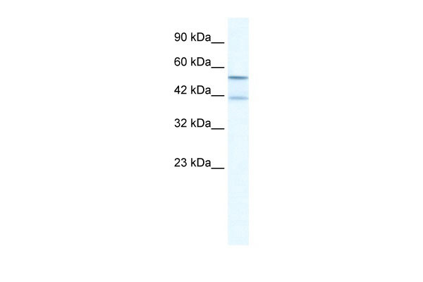
KCNK10 antibody - N-terminal region
Rabbit Polyclonal Antibody
- SPECIFICATION
- CITATIONS
- PROTOCOLS
- BACKGROUND

Application
| WB |
|---|---|
| Primary Accession | P57789 |
| Other Accession | NM_021161, NP_066984 |
| Reactivity | Human, Mouse, Rat, Rabbit, Zebrafish, Pig, Horse, Bovine, Dog |
| Predicted | Human, Mouse, Rabbit, Zebrafish, Pig, Chicken, Bovine |
| Host | Rabbit |
| Clonality | Polyclonal |
| Calculated MW | 59kDa |
| Gene ID | 54207 |
|---|---|
| Alias Symbol | TREK2, TREK-2, K2p10.1 |
| Other Names | Potassium channel subfamily K member 10, Outward rectifying potassium channel protein TREK-2, TREK-2 K(+) channel subunit, KCNK10, TREK2 |
| Format | Liquid. Purified antibody supplied in 1x PBS buffer with 0.09% (w/v) sodium azide and 2% sucrose. |
| Reconstitution & Storage | Add 100 ul of distilled water. Final anti-KCNK10 antibody concentration is 1 mg/ml in PBS buffer with 2% sucrose. For longer periods of storage, store at 20°C. Avoid repeat freeze-thaw cycles. |
| Precautions | KCNK10 antibody - N-terminal region is for research use only and not for use in diagnostic or therapeutic procedures. |
| Name | KCNK10 {ECO:0000303|PubMed:25766236, ECO:0000312|HGNC:HGNC:6273} |
|---|---|
| Function | K(+) channel that conducts voltage-dependent outward rectifying currents upon membrane depolarization. Voltage sensing is coupled to K(+) electrochemical gradient in an 'ion flux gating' mode where outward but not inward ion flow opens the gate. Converts to voltage-independent 'leak' conductance mode upon stimulation by various stimuli including mechanical membrane stretch, acidic pH, heat and lipids (PubMed:10880510, PubMed:25766236, PubMed:26919430, PubMed:38605031). Homo- and heterodimerizes to form functional channels with distinct regulatory and gating properties (PubMed:30573346). In trigeminal ganglia sensory neurons, the heterodimer of KCNK10/TREK-2 and KCNK18/TRESK inhibits neuronal firing and neurogenic inflammation by stabilizing the resting membrane potential at K(+) equilibrium potential as well as by regulating the threshold of action potentials and the spike frequency (By similarity). Permeable to other monovalent ions such as Rb(+) and Cs(+) (PubMed:26919430). |
| Cellular Location | Cell membrane {ECO:0000250|UniProtKB:Q8BUW1}; Multi-pass membrane protein |
| Tissue Location | [Isoform A]: Abundantly expressed in pancreas and kidney and to a lower level in brain, testis, colon, and small intestine. In brain, mainly expressed in cerebellum, occipital lobe, putamen, and thalamus. No expression is detected in amygdala and spinal cord. [Isoform C]: Abundantly expressed in brain. |

Thousands of laboratories across the world have published research that depended on the performance of antibodies from Abcepta to advance their research. Check out links to articles that cite our products in major peer-reviewed journals, organized by research category.
info@abcepta.com, and receive a free "I Love Antibodies" mug.

Provided below are standard protocols that you may find useful for product applications.
References
Gu,W., et al., (2002) J. Physiol. (Lond.) 539 (Pt 3), 657-668Reconstitution and Storage:For short term use, store at 2-8C up to 1 week. For long term storage, store at -20C in small aliquots to prevent freeze-thaw cycles.
If you have used an Abcepta product and would like to share how it has performed, please click on the "Submit Review" button and provide the requested information. Our staff will examine and post your review and contact you if needed.
If you have any additional inquiries please email technical services at tech@abcepta.com.
Ordering Information
Other Products
Shipping Information
