


 Foundational characteristics of cancer include proliferation, angiogenesis, migration, evasion of apoptosis, and cellular immortality. Find key markers for these cellular processes and antibodies to detect them.
 The SUMOplot™ Analysis Program predicts and scores sumoylation sites in your protein. SUMOylation is a post-translational modification involved in various cellular processes, such as nuclear-cytosolic transport, transcriptional regulation, apoptosis, protein stability, response to stress, and progression through the cell cycle.
 The Autophagy Receptor Motif Plotter predicts and scores autophagy receptor binding sites in your protein. Identifying proteins connected to this pathway is critical to understanding the role of autophagy in physiological as well as pathological processes such as development, differentiation, neurodegenerative diseases, stress, infection, and cancer.
> home > Products > Primary Antibodies > Antibody Collections > Zebrafish > Fnbp1 antibody - N-terminal region
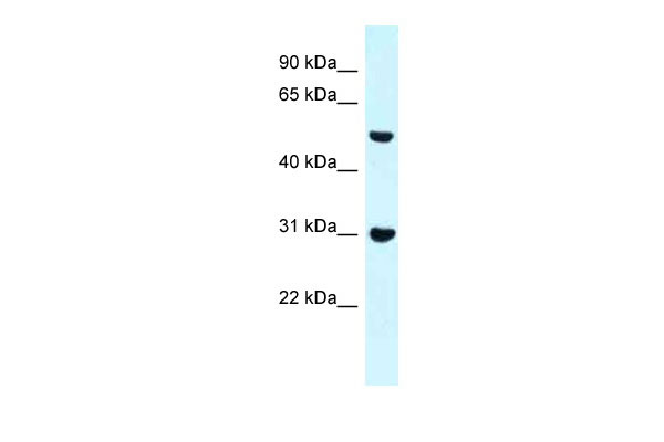
Fnbp1 antibody - N-terminal region
Rabbit Polyclonal Antibody
- SPECIFICATION
- CITATIONS
- PROTOCOLS
- BACKGROUND

Application
| WB |
|---|---|
| Primary Accession | Q80TY0 |
| Other Accession | NM_001177650, NP_001171121 |
| Reactivity | Human, Mouse, Rat, Rabbit, Zebrafish, Horse, Bovine, Dog |
| Predicted | Human, Mouse, Rat, Chicken, Bovine |
| Host | Rabbit |
| Clonality | Polyclonal |
| Calculated MW | 57kDa |
| Gene ID | 14269 |
|---|---|
| Alias Symbol | 1110057E06Rik, 2210010H06Rik, FBP1, Fbp17 |
| Other Names | Formin-binding protein 1, Formin-binding protein 17, Fnbp1, Fbp17, Kiaa0554 |
| Format | Liquid. Purified antibody supplied in 1x PBS buffer with 0.09% (w/v) sodium azide and 2% sucrose. |
| Reconstitution & Storage | Add 50 ul of distilled water. Final anti-Fnbp1 antibody concentration is 1 mg/ml in PBS buffer with 2% sucrose. For longer periods of storage, store at 20°C. Avoid repeat freeze-thaw cycles. |
| Precautions | Fnbp1 antibody - N-terminal region is for research use only and not for use in diagnostic or therapeutic procedures. |
| Name | Fnbp1 |
|---|---|
| Synonyms | Fbp17, Kiaa0554 |
| Function | Required to coordinate membrane tubulation with reorganization of the actin cytoskeleton during the late stage of clathrin-mediated endocytosis. Binds to lipids such as phosphatidylinositol 4,5-bisphosphate and phosphatidylserine and promotes membrane invagination and the formation of tubules. Also enhances actin polymerization via the recruitment of WASL/N-WASP, which in turn activates the Arp2/3 complex. Actin polymerization may promote the fission of membrane tubules to form endocytic vesicles. May act as a link between RND2 signaling and regulation of the actin cytoskeleton. May be required for the lysosomal retention of FASLG/FASL (By similarity). |
| Cellular Location | Cytoplasm. Cytoplasm, cytoskeleton. Cytoplasm, cell cortex. Lysosome. Cytoplasmic vesicle. Cell membrane; Peripheral membrane protein; Cytoplasmic side. Membrane, clathrin-coated pit Note=Enriched in cortical regions coincident with F-actin. Also localizes to endocytic vesicles and lysosomes. |
| Tissue Location | Expressed in brain and testis. |
Research Areas
Citations (0)

Thousands of laboratories across the world have published research that depended on the performance of antibodies from Abcepta to advance their research. Check out links to articles that cite our products in major peer-reviewed journals, organized by research category.
Submit your citation using an Abcepta antibody to
info@abcepta.com, and receive a free "I Love Antibodies" mug.
info@abcepta.com, and receive a free "I Love Antibodies" mug.

Application Protocols
Provided below are standard protocols that you may find useful for product applications.
Abcepta welcomes feedback from its customers.
If you have used an Abcepta product and would like to share how it has performed, please click on the "Submit Review" button and provide the requested information. Our staff will examine and post your review and contact you if needed.
If you have any additional inquiries please email technical services at tech@abcepta.com.
$ 389.00
Cat# AI11798
Ordering Information
United States
AlbaniaAustraliaAustriaBelgiumBosnia & HerzegovinaBrazilBulgariaCanadaCentral AmericaChinaCroatiaCyprusCzech RepublicDenmarkEstoniaFinlandFranceGermanyGreeceHong KongHungaryIcelandIndiaIndonesiaIrelandIsraelItalyJapanLatviaLithuaniaLuxembourgMacedoniaMalaysiaMaltaNetherlandsNew ZealandNorwayPakistanPolandPortugalRomaniaSerbiaSingaporeSlovakiaSloveniaSouth AfricaSouth KoreaSpainSwedenSwitzerlandTaiwanTurkeyUnited KingdomUnited StatesVietnamWorldwideOthers
USA Headquarters
(888) 735-7227 / (858) 622-0099 or (858) 875-1900
Other Products
Shipping Information
Domestic orders (in stock items)
Shipped out the same day. Orders placed after 1 PM (PST) will ship out the next business day.
International orders
Contact your local distributors
